. 2012 Aug 1;83(5):e661-7.
doi: 10.1016/j.ijrobp.2012.01.080. Epub 2012 May 7.

Esophageal toxicity from high-dose, single-fraction paraspinal stereotactic radiosurgery

Affiliations

Esophageal toxicity from high-dose, single-fraction paraspinal stereotactic radiosurgery

Brett W Cox et al. Int J Radiat Oncol Biol Phys. .

Abstract

Purpose: To report the esophageal toxicity from single-fraction paraspinal stereotactic radiosurgery (SRS) and identify dosimetric and clinical risk factors for toxicity.

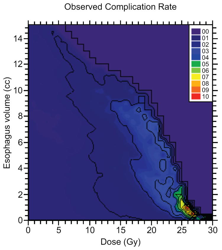
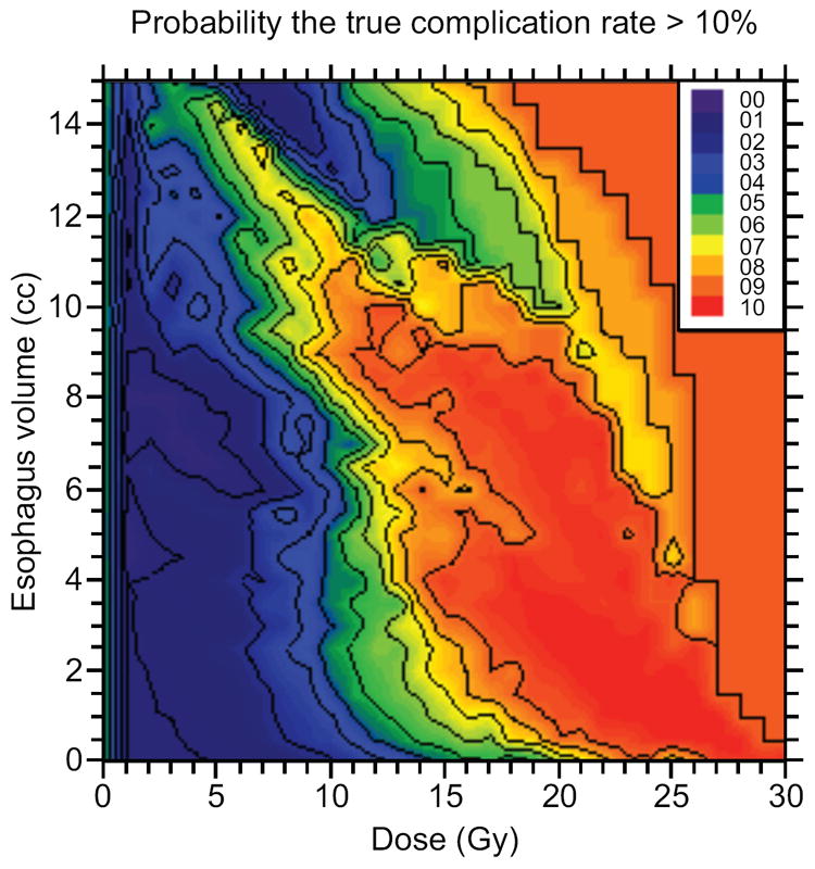
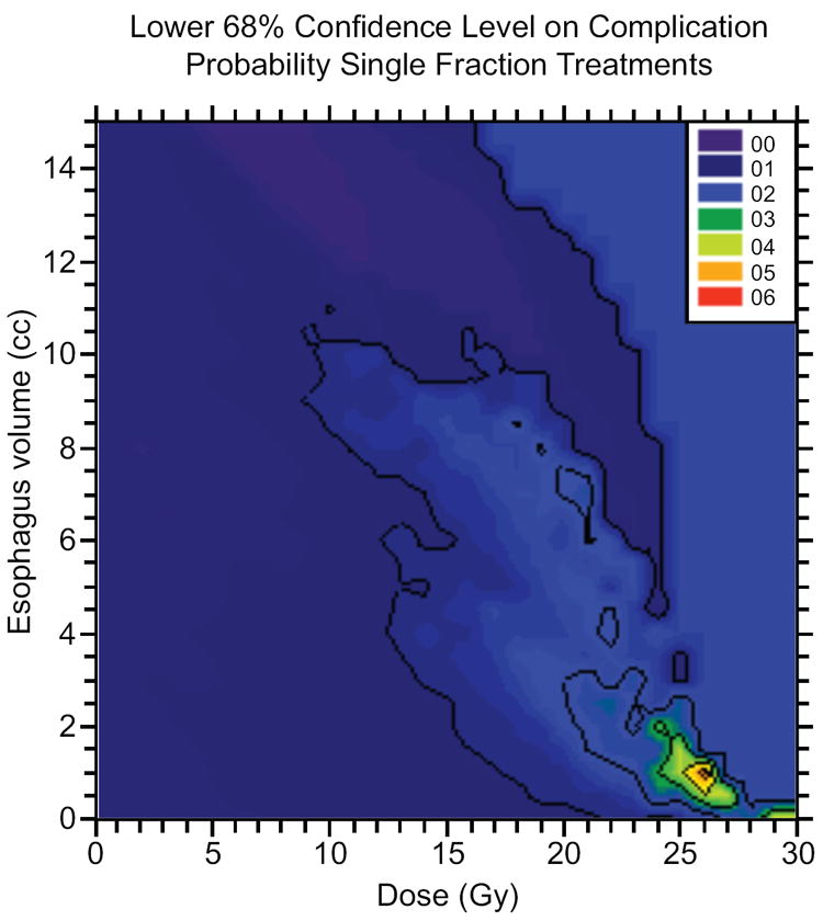
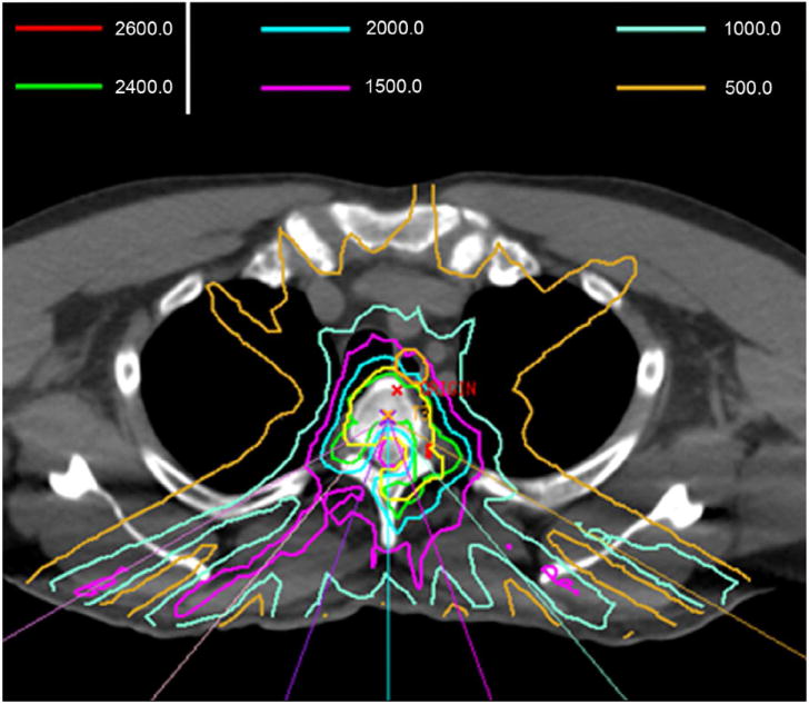
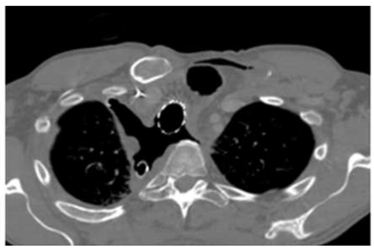
Methods and materials: A total of 204 spinal metastases abutting the esophagus (182 patients) were treated with high-dose single-fraction SRS during 2003-2010. Toxicity was scored using the National Cancer Institute Common Toxicity Criteria for Adverse Events, version 4.0. Dose-volume histograms were combined to generate a comprehensive atlas of complication incidence that identifies risk factors for toxicity. Correlation of dose-volume factors with esophageal toxicity was assessed using Fisher's exact test and logistic regression. Clinical factors were correlated with toxicity.

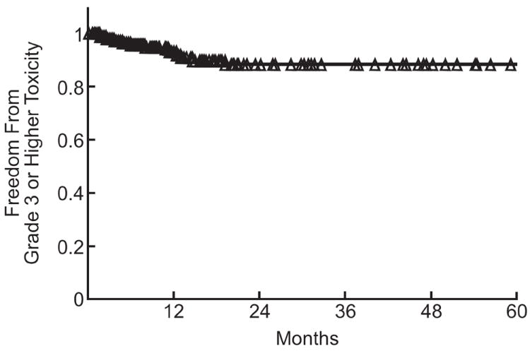
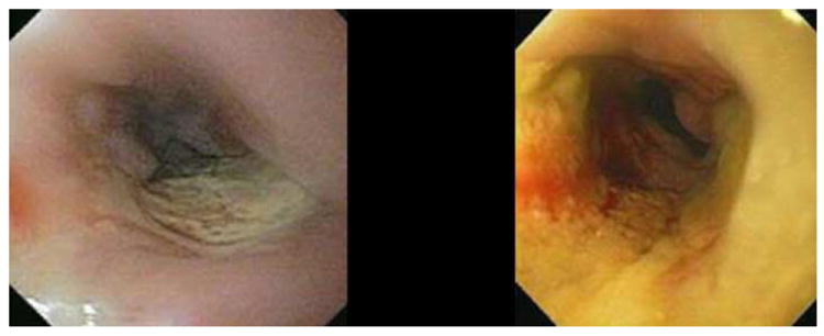
Results: The median dose to the planning treatment volume was 24 Gy. Median follow-up was 12 months (range, 3-81). There were 31 (15%) acute and 24 (12%) late esophageal toxicities. The rate of grade ≥3 acute or late toxicity was 6.8% (14 patients). Fisher's exact test resulted in significant median splits for grade ≥3 toxicity at V12 = 3.78 cm(3) (relative risk [RR] 3.7, P=.05), V15 = 1.87 cm(3) (RR 13, P=.0013), V20 = 0.11 cm(3) (RR 6, P=0.01), and V22 = 0.0 cm(3) (RR 13, P=.0013). The median split for D2.5 cm(3) (14.02 Gy) was also a significant predictor of toxicity (RR 6; P=.01). A highly significant logistic regression model was generated on the basis of D2.5 cm(3). One hundred percent (n = 7) of grade ≥4 toxicities were associated with radiation recall reactions after doxorubicin or gemcitabine chemotherapy or iatrogenic manipulation of the irradiated esophagus.

Conclusions: High-dose, single-fraction paraspinal SRS has a low rate of grade ≥3 esophageal toxicity. Severe esophageal toxicity is minimized with careful attention to esophageal doses during treatment planning. Iatrogenic manipulation of the irradiated esophagus and systemic agents classically associated with radiation recall reactions are associated with development of grade ≥4 toxicity.


Conflict of interest statement

Conflict of Interest Notification

Drs. Cox and Bilsky have no actual or potential conflicts of interest.

Figures

Fig. 1
Fig. 1
Actuarial analysis of grade ≥3 esophageal toxicity. The number of treated sites at risk for toxicity at 3 month intervals during the first 24 months was: 0 months, 204; 3 months, 165; 6 months, 142; 9 months, 122; 12 months, 102; 15 months, 85; 18 months, 64; 21 months, 50; 24months, 40.
Fig. 2
Fig. 2
a) Observed severe esophageal complication rate for DVHs passing over a given position in the dose-volume plane, plotted as a function of that position. b) Probability of a true esophageal complication rate >10% for DVHs passing over a point in the dose-volume plane for the esophagus, plotted at that point. C) Represents the lower 68% confidence level of complication probability for single-fraction treatments for DVHs passing over a point in the dose-volume plane for the esophagus. Esophageal doses of greater than 25 Gy to even small volumes of esophagus yields a high probability of esophageal complication. D) demonstrates the upper 68% confidence level for complication rate for DVHs passing over a given position in the dose/volume plane, plotted as a function of that position.
Fig. 2
Fig. 2
a) Observed severe esophageal complication rate for DVHs passing over a given position in the dose-volume plane, plotted as a function of that position. b) Probability of a true esophageal complication rate >10% for DVHs passing over a point in the dose-volume plane for the esophagus, plotted at that point. C) Represents the lower 68% confidence level of complication probability for single-fraction treatments for DVHs passing over a point in the dose-volume plane for the esophagus. Esophageal doses of greater than 25 Gy to even small volumes of esophagus yields a high probability of esophageal complication. D) demonstrates the upper 68% confidence level for complication rate for DVHs passing over a given position in the dose/volume plane, plotted as a function of that position.
Fig. 3
Fig. 3
Dose-response model for grade ≥3 esophagitis with single-fraction spine radiosurgery, from logistic regression using D 2.5 cm3. When the D 2.5 cm3 <14 Gy, there is a <5% probability of grade ≥3 esophageal toxicity, with a steep increase in toxicity at higher doses (p<0.01). For comparison, observed complication rates in quartiles in D 2.5 cm3 are plotted at the median value of the quartile. Black vertical lines, 68% confidence limits on the observed complication rates; horizontal lines, central 68% of the dose values in the quartile.
Figure 4
Figure 4
Representative grade 4 esophageal complication after spine radiosurgery showing the role of iatrogenic manipulation in development of high-grade toxicity. Patient was a 45-year-old man with oligometastatic renal cell carcinoma treated with 24 Gy to a symptomatic T3 lesion. a) shows representative isodose distributions. Esophageal planning constraints kept the D2.0 cm3 esophagus <15 Gy. The patient experienced grade 2 esophagitis at 4 months, and an esophagogastroduodenoscopy (EGD) showed a 3 cm nonbleeding ulcer (b) that was biopsied. Pain immediately worsened, and repeat EGD at 6 months showed increase in size, extent, and severity with superinfection (c). Biopsy and dilation was performed in the absence of stricture. Two weeks later the patient acutely developed a tracheoesophageal fistula (TEF) requiring multiple stent and repair procedures. d) A thoracic CT image demonstrating multiple stigmata of TEF formation, including tracheal and esophageal stents in place, pneumomediastinum, and soft-tissue defects. The patient died from distant progression of disease at 11 months.
Figure 4
Figure 4
Representative grade 4 esophageal complication after spine radiosurgery showing the role of iatrogenic manipulation in development of high-grade toxicity. Patient was a 45-year-old man with oligometastatic renal cell carcinoma treated with 24 Gy to a symptomatic T3 lesion. a) shows representative isodose distributions. Esophageal planning constraints kept the D2.0 cm3 esophagus <15 Gy. The patient experienced grade 2 esophagitis at 4 months, and an esophagogastroduodenoscopy (EGD) showed a 3 cm nonbleeding ulcer (b) that was biopsied. Pain immediately worsened, and repeat EGD at 6 months showed increase in size, extent, and severity with superinfection (c). Biopsy and dilation was performed in the absence of stricture. Two weeks later the patient acutely developed a tracheoesophageal fistula (TEF) requiring multiple stent and repair procedures. d) A thoracic CT image demonstrating multiple stigmata of TEF formation, including tracheal and esophageal stents in place, pneumomediastinum, and soft-tissue defects. The patient died from distant progression of disease at 11 months.

References

    1. Ryu SI, Chang SD, Kim DH, et al. Image-guided hypo-fractionated stereotactic radiosurgery to spinal lesions. Neurosurgery. 2001;49:838–846. - PubMed
    1. Yamada Y, Bilsky MH, Lovelock DM, et al. High-dose, single-fraction image-guided intensity-modulated radiotherapy for metastatic spinal lesions. Int J Radiat Oncol Biol Phys. 2008;71:484–490. - PubMed
    1. Gerszten PC, Burton SA, Ozhasoglu C, et al. Radiosurgery for spinal metastases: clinical experience in 500 cases from a single institution. Spine (Phila Pa 1976) 2007;32:193–199. - PubMed
    1. Gibbs IC, Kamnerdsupaphon P, Ryu MR, et al. Image-guided robotic radiosurgery for spinal metastases. Radiother Oncol. 2007;82:185–190. - PubMed
    1. Katagiri H, Takahashi M, Inagaki J, et al. Clinical results of nonsurgical treatment for spinal metastases. Int J Radiat Oncol Biol Phys. 1998;42:1127–1132. - PubMed

Publication types