Review
. 2012 Jun;2(3):264-75.
doi: 10.1016/j.coviro.2012.04.004. Epub 2012 May 7.

SARS coronavirus pathogenesis: host innate immune responses and viral antagonism of interferon

Affiliations
Review

SARS coronavirus pathogenesis: host innate immune responses and viral antagonism of interferon

Allison L Totura et al. Curr Opin Virol. 2012 Jun.

Abstract

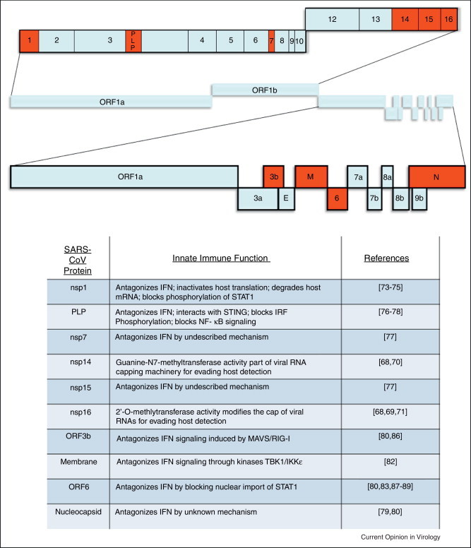
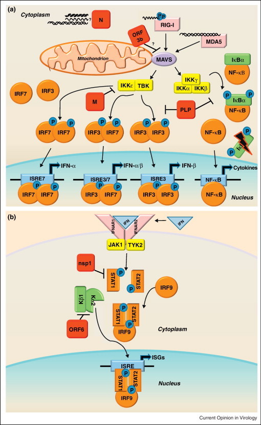
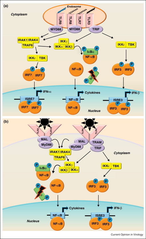
SARS-CoV is a pathogenic coronavirus that emerged from a zoonotic reservoir, leading to global dissemination of the virus. The association SARS-CoV with aberrant cytokine, chemokine, and Interferon Stimulated Gene (ISG) responses in patients provided evidence that SARS-CoV pathogenesis is at least partially controlled by innate immune signaling. Utilizing models for SARS-CoV infection, key components of innate immune signaling pathways have been identified as protective factors against SARS-CoV disease, including STAT1 and MyD88. Gene transcription signatures unique to SARS-CoV disease states have been identified, but host factors that regulate exacerbated disease phenotypes still remain largely undetermined. SARS-CoV encodes several proteins that modulate innate immune signaling through the antagonism of the induction of Interferon and by avoidance of ISG effector functions.


Figures

Figure 1
Figure 1
The SARS-CoV genome and functions of SARS-CoV innate immune antagonists. (a) The typical coronavirus genome size is quite large in comparison to many other positive-sense RNA viruses; within the SARS-CoV genome of 29.7 kB at least ten genes with potential functions that modulate innate immunity have been characterized (highlighted here in red). Like other members of the viral family Coronaviridae, SARS-CoV has a positive-sense, single-stranded RNA genome that is amenable to manipulation using reverse genetic techniques [90]. In SARS-CoV the first open reading frame (ORF) encodes the 16 nonstructural proteins that make up the viral replicase, while the ensuing ORFs encode four structural proteins that compose the virion, as well as eight accessory proteins. The SARS-CoV accessory proteins share no homology to the accessory proteins of other human coronaviruses, and while dispensable for replication in vitro, encode functions that probably impact viral pathogenesis in vivo [91]. While SARS-CoV was a novel virus not previously recognized before the 2002 outbreak, other coronaviruses have been associated with disease in humans. Coronaviruses known to infect humans include HCoV-HKU1, HCoV-OC43, HCoV-229E, and HCoV-NL63, which also cause respiratory infections but are generally much less severe than SARS [92]. (b) Transcription and subsequent signaling of Interferon is vital for activating the antiviral response in host cells. Because of this, many viruses (including SARS-CoV) encode proteins that antagonize the IFN response to viral infection. Of the SARS-CoV viral proteins listed here, eight have been identified as Interferon antagonists and two have been implicated in the viral RNA capping machinery.
Figure 2
Figure 2
(a) RLR family of innate immune receptors induce Type I interferon. The family of RIG-I Like Receptors (RLRs) contains three cytosolic RNA helicases that recognize non-self RNA species resulting from viral replication [93]. The two signaling sensors within the RLR family are retinoic acid-inducible gene I (RIG-I) and melanoma differentiation associated factor 5 (MDA5). The third RLR, laboratory of genetics and physiology 2 (LGP2, not shown), facilitates recognition of viral PAMPs by RIG-I and MDA5, but is dispensable for their signaling [94]. RIG-I recognizes primarily 5′ppp-RNA molecules with secondary motifs of dsRNA or ssRNA of short length [95, 96]. MDA5 recognizes longer dsRNA motifs than RIG-I [97]. Following binding of viral RNAs, RIG-I and MDA5 interact with the mitochondrial membrane bound adaptor molecule MAVS (mitochondrial antiviral signaling protein, also referred to as IPS-1, VISA, or CARDIF) to transduce the signal via complexes of kinases: the IKKɛ/TBK1 complex and the IKKα/IKKβ/IKKγ complex. The IKKɛ/TBK1 kinases phosphorylate the transcription factors IRF3 and IRF7, which then form homodimers or heterodimers. Upon dimerization, the transcription factors enter the nucleus to initiate transcription of Type I IFNs (IFN-α and IFN-β). While IRF3 is nearly ubiquitously expressed in cells, IRF7 is an ISG typically expressed at low levels, so it is thought that IRF3 mediates transcription of the majority of early IFN expression. The IKKα/IKKβ/IKKγ kinases phosphorylate IκBα, targeting this repressor protein of NF-κB for degradation. Activation of NF-κB leads to transcription of proinflammatory cytokines, and NF-kB mediated transcription has also been linked to the pathogenesis of ARDS [46]. SARS-CoV encodes proteins that antagonize RLR family signaling, shown here in red. (b) Interferon signals through the JAK-STAT pathway to induce interferon stimulated genes. The secretion of IFN-α and IFN-β molecules from an infected cell leads to an autocrine and paracrine signaling through the IFNαβ Receptor (composed of the IFNAR1 and IFNAR2 subunits) resulting in the activation of the JAK-STAT pathway. The JAK/TYK2 kinases phosphorylate the transcription factors STAT1 and STAT2, which form heterodimers complexed with IRF9. The STAT complex translocates to the nucleus leading to the transcription of Interferon Stimulated Genes (ISGs) that establish an antiviral state in the cell. Because neighboring cells can receive IFN stimulation before infection, it is a crucial pathway to preventing viral spread in the host. SARS-CoV also encodes proteins that antagonize the JAK-STAT pathway, shown here in red. Mice deficient in STAT1 showed an increased susceptibility to SARS-CoV infection [98]. Although there were no differences in mice deficient in IFN receptors, STAT1−/− mice showed increased weight loss, viral titer, and lung pathology compared to wild type over the course of MA15-SARS-CoV infection, demonstrating that STAT has important IFN independent role in SARS-CoV infection [80]. Severe lung pathology in STAT1−/− mice infected with MA15-SARS-CoV was associated with the infiltration of immune cells and fibrotic lung response. The STAT1−/− dependent prolonged expression of inflammatory cytokines (IL-1, IL-6, IL-10, IL-12, and TNFα) and chemokines (CCL2, CCL3, CCL4, CCL7, and CCL20), could be a transcriptional regime responsible for fibrotic phenotypes within the lungs. Additionally, ISG responses were significantly lower in STAT1−/− mice compared to wild type or IFNAR−/− mice, leading to the conclusion that STAT1 dependent, IFNAR1 independent ISG expression was protective in these mice [80]. It remains unclear how STAT1 controls ISGs independent of IFNAR expression, or which ISGs have important potential roles in SARS-CoV pathogenesis.
Figure 3
Figure 3
Pathogen associated molecular pattern sensing by Toll-Like Receptors. (a) In the endosomal compartment, TLRs recognize viral nucleic acid PAMPs: TLR3 recognizes dsRNAs, TLR7/8 recognizes ssRNAs, and TLR9 recognizes CpG DNA motifs. (b) On the surface of cells, TLR2 and TLR4 are known to recognize viral glycoproteins [47, 99]. TLR2/6 heterodimers help to activate the innate immune response to RSV, though the viral PAMP recognized has not been determined [100]. TLR1/2 heterodimers have been shown to recognize viral glycoproteins, though their potential role in respiratory virus infection has not been determined [99]. While there are many TLRs that recognize viral PAMPs, they signal through common adaptor molecules, including MyD88, MAL, TRAM, and TRIF. The TLR adaptor molecules signal through the IKKɛ/TBK1 complex and the IKKα/IKKβ/IKKγ complex similarly to RLRs, but can also recruit an IRAK-1/IRAK4/TRAF6 complex capable of activating the transcription factors IRF3, IRF7, and NF-κB. Activation of these transcription factors leads to the transcription of Type I IFNs and proinflammatory cytokines. Due to the considerable crosstalk between TLR and RLR signaling, it is difficult to discriminate between transcriptional products generated by the two sensor families, but it is likely that both play an important role in the innate immune response to SARS-CoV infection.

References

    1. Drosten C., Gunther S., Preiser W., van der Werf S., Brodt H.-R., Becker S., Rabenau H., Panning M., Kolesnikova L., Fouchier R.A.M. Identification of a novel coronavirus in patients with severe acute respiratory syndrome. New England Journal of Medicine. 2003;348:1967–1976. - PubMed
    1. Ksiazek T.G., Erdman D., Goldsmith C.S., Zaki S.R., Peret T., Emery S., Tong S., Urbani C., Comer J.A., Lim W. A novel coronavirus associated with severe acute respiratory syndrome. New England Journal of Medicine. 2003;348:1953–1966. - PubMed
    1. Li W., Moore M.J., Vasilieva N., Sui J., Wong S.K., Berne M.A., Somasundaran M., Sullivan J.L., Luzuriaga K., Greenough T.C. Angiotensin-converting enzyme 2 is a functional receptor for the SARS coronavirus. Nature. 2003;426:450–454. - PMC - PubMed
    1. Hui D.S.C., Chan M.C.H., Wu A.K., Ng P.C. Severe acute respiratory syndrome (SARS): epidemiology and clinical features. Postgraduate Medical Journal. 2004;80:373–381. - PMC - PubMed
    1. World Health Organization: Summary of probable SARS cases with onset of illness from 1 November 2002 to 31 July 2003 on World Wide Web URL: http://www.who.int/csr/sars/country/table2004_04_21/en/index.html.

Publication types

MeSH terms