. 2012 May 9;4(133):133ra56.
doi: 10.1126/scitranslmed.3003713.

Survivin is a therapeutic target in Merkel cell carcinoma

Affiliations

Survivin is a therapeutic target in Merkel cell carcinoma

Reety Arora et al. Sci Transl Med. .

Abstract

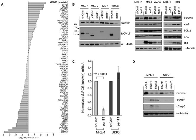
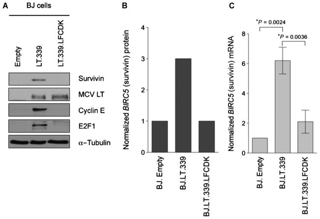
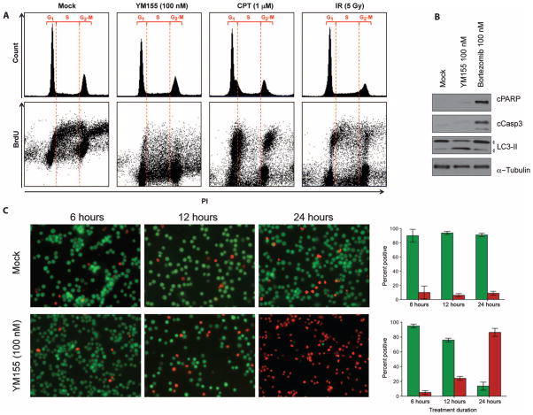
Merkel cell polyomavirus (MCV) causes ~80% of primary and metastatic Merkel cell carcinomas (MCCs). By comparing digital transcriptome subtraction deep-sequencing profiles, we found that transcripts of the cellular survivin oncoprotein [BIRC5a (baculoviral inhibitor of apoptosis repeat-containing 5)] were up-regulated sevenfold in virus-positive compared to virus-negative MCC tumors. Knockdown of MCV large T antigen in MCV-positive MCC cell lines decreased survivin mRNA and protein expression. Exogenously expressed MCV large T antigen increased survivin protein expression in non-MCC primary cells. This required an intact retinoblastoma protein-targeting domain that activated survivin gene transcription as well as expression of other G(1)-S-phase proteins including E2F1 and cyclin E. Survivin expression is critical to the survival of MCV-positive MCC cells. A small-molecule survivin inhibitor, YM155, potently and selectively initiates irreversible, nonapoptotic, programmed MCV-positive MCC cell death. Of 1360 other chemotherapeutic and pharmacologically active compounds screened in vitro, only bortezomib (Velcade) was found to be similarly potent, but was not selective in killing MCV-positive MCC cells. YM155 halted the growth of MCV-positive MCC xenograft tumors and was nontoxic in mice, whereas bortezomib was not active in vivo and mice displayed serious morbidity. Xenograft tumors resumed growth once YM155 treatment was stopped, suggesting that YM155 may be cytostatic rather than cytotoxic in vivo. Identifying the cellular pathways, such as those involving survivin, that are targeted by tumor viruses can lead to rapid and rational identification of drug candidates for treating virus-induced cancers.


Conflict of interest statement

Competing interests: Y.C. and P.S.M. have submitted a patent application U.S. 12/808,042 entitled “Methods to diagnose and immunize against the virus causing human Merkel cell carcinoma.” This manuscript is the basis of a pending patent application and, if issued, Y.C. and P.S.M. may be entitled to receive royalties under University of Pittsburgh policy.

Figures

Fig. 1
Fig. 1
Survivin oncoprotein mRNA expression is increased in MCV-positive MCC. (A) Digital transcriptome subtraction comparison of 64 genes involved in programmed cell death and cell cycle regulation showed that survivin (BIRC5) mRNA transcripts (highlighted) were sevenfold higher in an MCV-positive compared with an MCV-negative MCC cDNA library. The relative expression of genes was normalized to total sequence reads for each MCC library (also see table S1). (B) MCV T antigen is required for survivin expression. Lentiviral MCV T antigen exon 1 knockdown (panT1) decreased survivin protein expression (left) but did not alter XIAP, BCL-2, BAX, or p53 protein levels (right) in four MCV-positive MCC cell lines: MKL-1, MKL-2, MS-1, and WaGa. UISO, MCV-negative cell line; shCntrl, scrambled shRNA control lentivirus; LT, large T antigen. (C) MCV T antigen is required for survivin transcription. Survivin mRNA levels were reduced in the MKL-1 but not the UISO cell line after T antigen knockdown, indicating that T antigen activates survivin transcription. Survivin mRNA was measured by qRT-PCR and normalized to β-actin mRNA. The experiments were performed in triplicate and repeated twice (mean ± SEM, two-tailed t test). (D) Survivin expression is required for MCV-positive MCC tumor cell survival. Survivin was targeted for knockdown using two shRNA lentiviral vectors, shsur1 and shsur2, in MKL-1 and UISO cells. MKL-1 cells initiate apoptosis after survivin knockdown, with increased expression of cleaved PARP (cPARP) and caspase 3 (cCasp3), whereas UISO cells are resistant to survivin knockdown-induced apoptosis. α-Tubulin is used as a loading control.
Fig. 2
Fig. 2
MCV large T antigen isoform induces survivin oncoprotein expression in human BJ cells by targeting RB. (A) BJ cells were transduced with either empty vector, a tumor-derived large T antigen cDNA (LT.339), or a large T antigen cDNA with an inactive RB binding domain (LT.339LFCDK). Immunoblotting reveals that MCV LT.339 induces survivin expression but LT.339LFCDK does not. A similar pattern is seen for other S-phase cell cycle proteins such as E2F1 and cyclin E that are also transcriptionally repressed by RB. (B) LI-COR quantitative immunoblotting for survivin in (A), normalized to α-tubulin (arbitrary units). (C) Survivin mRNA levels increased in BJ cells expressing LT.339 protein but not in cells expressing the RB1 binding mutant LT.339LFCDK. BJ cells expressing either empty vector, LT.339, or LT.339LFCDK were serum-starved for 48 hours and then harvested for RNA. Survivin mRNA was measured by qRT-PCR and normalized to β-actin mRNA. The experiments were performed three times in duplicate (mean ± SEM, two-tailed t test).
Fig. 3
Fig. 3
The survivin promoter inhibitor YM155 inhibits MCV-positive MCC cell line growth. (A) Dose-dependent growth curves at 48 hours for YM155-treated cell lines. MCV-negative MCC13, BJ, and BJhTERT cells showed relative resistance to YM155 treatment, whereas all MCV-positive cell lines (MKL-1, MKL-2, MS-1, and WaGa) were sensitive to YM155. MCV-negative UISO and MCC26 cell lines had intermediate sensitivity to YM155. (B) Trypan blue vital dye exclusion assay showed dose-dependent cell killing at 48 hours for MKL-1 cells (blue bars), whereas UISO cells (red bars) are relatively less sensitive and BJ cells (green bars) are resistant to YM155. (C) Dose-dependent decrease in MKL-1 cell survivin protein expression after 12 hours of YM155 treatment.
Fig. 4
Fig. 4
Cell death phenotype of YM155-treated MCV-positive MCC. (A) Cell cycle analysis reveals that YM155-treated cells do not undergo mitotic catastrophe. MKL-1 cells were treated with DMSO, YM155 (100 nM), camptothecin (CPT, 1 μM), or 5-Gy γ-irradiation (IR). They were stained for propidium iodide (PI) and BrdU and harvested 12 hours later. Upper panel shows cell cycle profiles, in which no evidence of G2-M pileup is seen (a marker for mitotic catastrophe) after YM155 treatment. Bottom panel shows the corresponding propidium iodide versus BrdU uptake for these same cells showing new DNA synthesis for cells in G1, S, and G2-M. Mock-treated cells have an inverted U-shaped curve showing BrdU incorporation during S-phase DNA synthesis. Both camptothecin and γ-irradiation primarily reduce S-phase BrdU incorporation, consistent with checkpoint activation. In contrast, YM155 reduces all early DNA synthesis as measured by BrdU incorporation. (B) YM155 induces nonapoptotic cell death associated with autophagy in MKL-1 cells. MKL-1 cells were treated with DMSO, YM155 (100 nM), or bortezomib (100 nM) and immunoblotted for cleaved PARP (cPARP), cleaved caspase 3 (cCasp3), LC3-II, and α-tubulin. In contrast to YM155, the proteasome inhibitor bortezomib activates MKL-1 cell apoptosis (also see fig. S2). (C) YM155 treatment initiates programmed cell death within 12 to 24 hours after treatment. MKL-1 cells were costained with CFDA (green, live) and propidium iodide (red, dead). Column graphs (right panel) represent mean and range of percent CFDA-positive (green) and percent propidium iodide–positive cells (red).
Fig. 5
Fig. 5
YM155 inhibits growth of human MKL-1 MCC xenografts in NOD-SCIDγ mice. (A) MKL-1 xenograft tumors were stained with hematoxylin and eosin (H&E), and for MCV large T antigen and cytokeratin 20 (CK20) expression (magnification, ×40). (B) MKL-1 xenograft survival curves after drug treatment. Mice were subcutaneously injected with 20 million MKL-1 cells and assigned to a 3-week drug treatment after tumors became palpable. No significant difference was found between saline and bortezomib treatment. Tumor progression was significantly delayed by YM155, with none of the YM155-treated mice dying during treatment (up to day 19) compared to 23 of 31 (74%) saline-treated and 14 of 21 (67%) bortezomib-treated mice (P < 0.0001, log-rank test). Tumor progression recurred for all YM155-treated mice once treatment was stopped. (C) Piecewise linear hierarchical Bayesian model for tumor volumes in treated mice (59) (see the Supplementary Materials for details). Colored lines show estimated central population tumor volumes, with shaded regions representing 95% credible intervals. Actual tumor volumes (gray lines) for each mouse are shown for comparison. YM155 treatment retards tumor growth compared to saline or bortezomib treatment. (D) Table showing day of termination and tumor volumes for MS-1 and UISO xenograft mice treated with YM155 or saline.

References

    1. Allen PJ, Bowne WB, Jaques DP, Brennan MF, Busam K, Coit DG. Merkel cell carcinoma: Prognosis and treatment of patients from a single institution. J Clin Oncol. 2005;23:2300–2309. - PubMed
    1. Veness M, Foote M, Gebski V, Poulsen M. The role of radiotherapy alone in patients with Merkel cell carcinoma: Reporting the Australian experience of 43 patients. Int J Radiat Oncol Biol Phys. 2010;78:703–709. - PubMed
    1. Eng TY, Boersma MG, Fuller CD, Goytia V, Jones WE, III, Joyner M, Nguyen DD. A comprehensive review of the treatment of Merkel cell carcinoma. Am J Clin Oncol. 2007;30:624–636. - PubMed
    1. Lemos B, Nghiem P. Merkel cell carcinoma: More deaths but still no pathway to blame. J Invest Dermatol. 2007;127:2100–2103. - PubMed
    1. Hodgson NC. Merkel cell carcinoma: Changing incidence trends. J Surg Oncol. 2005;89:1–4. - PubMed

Publication types

MeSH terms

LinkOut - more resources