. 2012 Aug 30;87(2):51.
doi: 10.1095/biolreprod.111.096511. Print 2012 Aug.

Peripubertal vitamin D(3) deficiency delays puberty and disrupts the estrous cycle in adult female mice

Affiliations

Peripubertal vitamin D(3) deficiency delays puberty and disrupts the estrous cycle in adult female mice

Cary L Dicken et al. Biol Reprod. .

Abstract

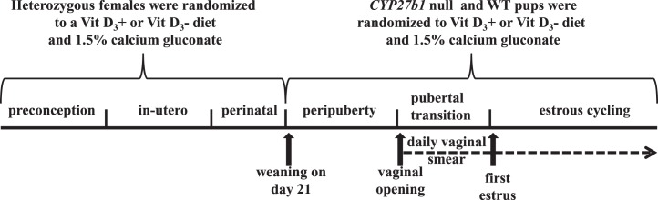
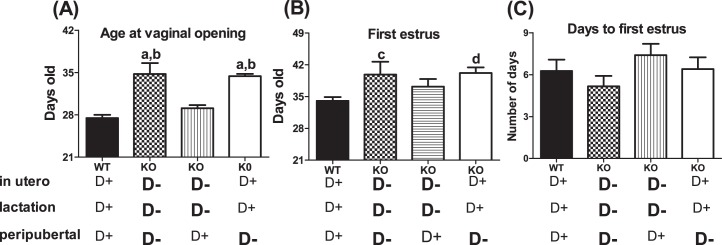
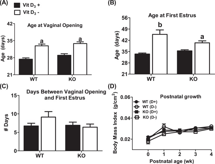
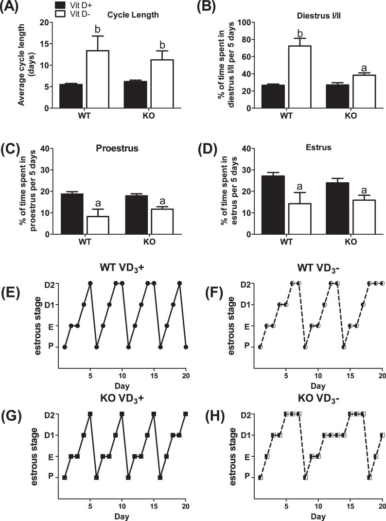
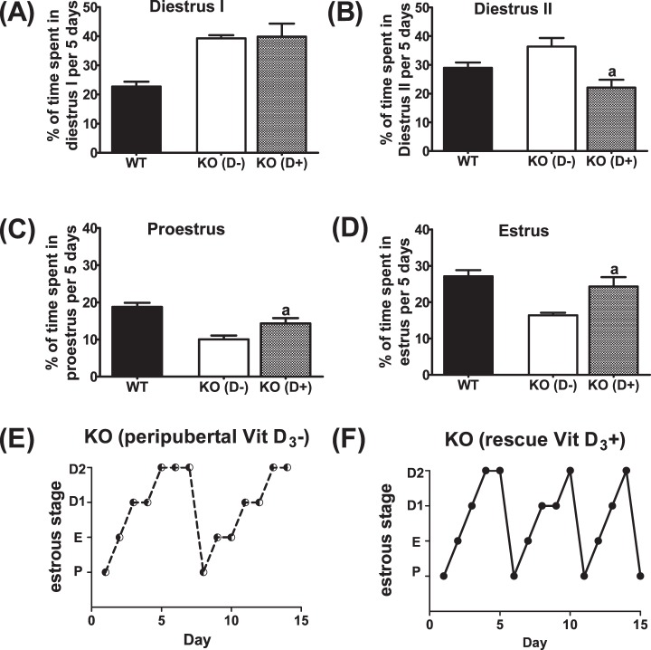
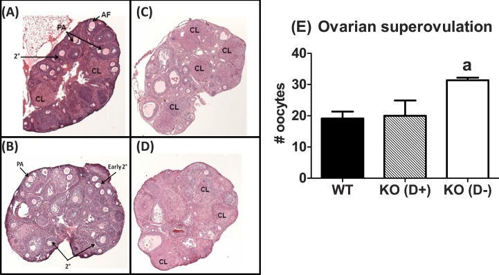
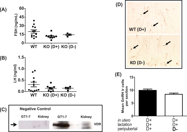
The mechanism(s) by which vitamin D(3) regulates female reproduction is minimally understood. We tested the hypothesis that peripubertal vitamin D(3) deficiency disrupts hypothalamic-pituitary-ovarian physiology. To test this hypothesis, we used wild-type mice and Cyp27b1 (the rate-limiting enzyme in the synthesis of 1,25-dihydroxyvitamin D(3)) null mice to study the effect of vitamin D(3) deficiency on puberty and reproductive physiology. At the time of weaning, mice were randomized to a vitamin D(3)-replete or -deficient diet supplemented with calcium. We assessed the age of vaginal opening and first estrus (puberty markers), gonadotropin levels, ovarian histology, ovarian responsiveness to exogenous gonadotropins, and estrous cyclicity. Peripubertal vitamin D(3) deficiency significantly delayed vaginal opening without affecting the number of GnRH-immunopositive neurons or estradiol-negative feedback on gonadotropin levels during diestrus. Young adult females maintained on a vitamin D(3)-deficient diet after puberty had arrested follicular development and prolonged estrous cycles characterized by extended periods of diestrus. Ovaries of vitamin D(3)-deficient Cyp27b1 null mice responded to exogenous gonadotropins and deposited significantly more oocytes into the oviducts than mice maintained on a vitamin D(3)-replete diet. Estrous cycles were restored when vitamin D(3)-deficient Cyp27b1 null young adult females were transferred to a vitamin D(3)-replete diet. This study is the first to demonstrate that peripubertal vitamin D(3) sufficiency is important for an appropriately timed pubertal transition and maintenance of normal female reproductive physiology. These data suggest vitamin D(3) is a key regulator of neuroendocrine and ovarian physiology.


Figures

FIG. 1.
FIG. 1.
Timeline of experimental events. All experiments were carried out with mice born from heterozygote matings in which the dams received a Vit D3+ or Vit D3− diet. WT and Cyp27b1 null mice female pups were weaned at 21 days and randomized to either a Vit D3+ or a Vit D3− diet supplemented with calcium gluconate. WT (+/+) mice that received a Vit D3+ diet throughout gestation, lactation, and weaning served as controls. All mice were inspected daily for vaginal opening. After vaginal opening, vaginal lavage was performed daily at 1500 h for a minimum of 7 wk, and estrous cycle length and estrous stage frequency were determined with vaginal smears.
FIG. 2.
FIG. 2.
Peripubertal 1,25-(OH)2 vitamin D3 deficiency delays puberty. A) Age at vaginal opening (VO) in WT (Cyp27b1+/+) mice fed a Vit D3+ diet throughout pregnancy (in utero) and lactation (perinatal period) and peripubertal transition and in Cyp27b1 null female mice subjected to a Vit D3− diet in utero and perinatal period and peripubertal transition, or in utero and perinatal period, or only during the peripubertal transition. B) Age at first estrus in WT mice fed a D+ diet in utero and perinatal period and peripubertal transition and in Cyp27b1 null female mice subjected to D− diet in utero and perinatal period and peripubertal transition or in utero and perinatal period, or only during the peripubertal transition. C) Number of days between VO and first estrus in WT mice fed a D+ diet in utero and perinatal period and peripubertal transition and in Cyp27b1 null female mice subjected to D diet in utero and perinatal period and peripubertal transition, or in utero and perinatal period, or only during the peripubertal transition (n = 8–15); aP < 0.0001 vs. WT; bP < 0.0001 vs. in utero and lactation; cP < 0.05 vs. WT; dP < 0.01 vs. WT.
FIG. 3.
FIG. 3.
Mice fed a vitamin D3-deficient diet during the prepubertal period have a delayed pubertal onset but normal developmental growth curves. A) Age at vaginal opening. B) Age at first estrus. C) Days between vaginal opening and first estrus. D) Developmental growth curves of mice. KO denotes Cyp27b1 null mice. WT denotes Cyp27b1+/+ mice. Vit D3+ or D+ denotes mice fed a Vit D3-sufficient diet before and after weaning. Vit D3− or D− denotes mice supplemented with calcium gluconate and fed a Vit D3-deficient diet after weaning (n = 8–15). aP < 0.01 vs. Vit D3+; bP < 0.0001 vs. Vit D3+
FIG. 4.
FIG. 4.
Vitamin D3 deficiency extends the estrous cycle by increasing time spent in diestrus. A) Average cycle length in WT and Cyp27b1 null mice fed a Vit D3+ or Vit D3− diet during the peripubertal period. Average percentage of time spent in diestrus I/II (B), proestrus (C), and estrus (D) in WT and Cyp27b1 null mice fed a Vit D3+ diet or Vit D3− diet during the prepubertal period and into early adulthood (n = 8–15); aP < 0.05 vs. Vit D3+ diet; bP < 0.001 vs. Vit D3+ diet. E) Representative estrous cycle of WT mouse fed Vit D3+ diet throughout gestation and lactation and after weaning. F) Representative estrous cycle of WT mouse fed Vit D3+ diet throughout gestation and lactation and weaned onto a Vit D3− diet. G) Representative estrous cycle of Cyp27b1 mouse fed Vit D3+ diet throughout gestation and lactation and after weaning. H) Representative estrous cycle of Cyp27b1 mouse fed Vit D3+ diet throughout gestation and lactation and weaned onto a Vit D3− diet. P, proestrus; E, estrus; D1, diestrus 1; D2, diestrus 2.
FIG. 5.
FIG. 5.
Vitamin D3 deficiency reversibly disrupts the estrous cycle. Average percentage of time spent in diestrus I (A), diestrus II (B), proestrus (C), and estrus (D) when Vit D3Cyp27b1 mice (KO [D-]) were switched to a Vit D3+ (D+) diet. WT (Vit D3+) mice described in Figure 4 were included for comparison. aP < 0.03 vs. WT and KO (D+) mice; n = 6. E) Representative estrous cycle of Cyp27b1 mouse fed Vit D3+ diet throughout gestation and lactation and weaned onto a Vit D3− diet. F) Representative estrous cycle of the same Cyp27b1 mouse transferred to a Vit D3+ diet for 4 wk. P, proestrus; E, estrus; D1, diestrus 1; D2, diestrus 2. (n = 6).
FIG. 6.
FIG. 6.
Diet-induced vitamin D3 deficiency is associated with a robust response to superovulation with exogenous gonadotropins. Representative photomicrograph (original magnification ×40) of WT (top) and Cyp27b1 null mice fed a Vit D3-deficient diet during the peripubertal period (bottom), injected with saline or superovulated with eCG plus hCG. A) WT mice injected with saline. B) Cyp27b1 null mice fed a Vit D3− diet, injected with saline. C) WT mice fed a Vit D3+ diet, injected with eCG and hCG. D) Cyp27b1 null mice fed a Vit D3− diet, injected with eCG and hCG. E) Number of oocytes deposited into the oviduct of WT and CYP27b1 null (KO) mice fed a Vit D3+ diet and Cyp27b1 null mice fed a Vit D3− diet after superovulation with eCG and hCG. AF, antral follicle; CL, corpus luteum; PA, early preantral; 2°, secondary. aP = 0.02 vs. WT and KO mice fed a Vit D3+ diet during the peripubertal transition (n = 4–6).
FIG. 7.
FIG. 7.
Effects of vitamin D3 on serum gonadotropin levels during mice in diestrus, VDR expression in GT1-7, and density of hypothalamic GnRH neurons. A) Serum FSH in reproduction-aged WT and Cyp27b1 null mice fed a vitamin D3-sufficient diet (KO [D+]) or a D3-deficient diet (KO [D−]) (n = 4–12). B) Serum LH in reproduction-aged WT and Cyp27b1 null mice fed a vitamin D3-sufficient diet (KO [D+]) or D3-deficient diet (KO [D−]) (n = 4–12). C) Western blot showing VDR in cell lysates from GT1-7 neurons and kidney. The positive control is WT mouse kidney, and negative control is GT1-7 and kidney lysate without primary VDR antibody. D) Representative sections of single-label immunohistochemistry (original magnification ×40) showing GnRH neurons (brown cytoplasm) in WT Vit D3+ and Cyp27b1 null Vit D3− mice. Arrows indicate GnRH immunoreactive neurons (n = 4). E) Average number of GnRH neuron numbers per hypothalamic section (means ± SEM) in WT Vit D3+ and Cyp27b1 null Vit D3− mice. Hypothalamic sections reviewed corresponded to plates 25–32 of the Paxinos and Watson mouse atlas [49] and the hypothalamic region between the organum vasculosum of lamina terminalis and the medial POA (n = 4).

References

    1. Christakos S, Barletta F, Huening M, Dhawan P, Liu Y, Porta A, Peng X. Vitamin D target proteins: function and regulation. J Cell Biochem 2003; 88 (2): 238 244 - PubMed
    1. Christakos S, Dhawan P, Liu Y, Peng X, Porta A. New insights into the mechanisms of vitamin D action. J Cell Biochem 2003; 88 (4): 695 705 - PubMed
    1. Christakos S, Dhawan P, Peng X, Obukhov AG, Nowycky MC, Benn BS, Zhong Y, Liu Y, Shen Q. New insights into the function and regulation of vitamin D target proteins. J Steroid Biochem Mol Biol 2007; 103 (3–5): 405 410 - PMC - PubMed
    1. Eyles D, Almeras L, Benech P, Patatian A, Mackay-Sim A, McGrath J, Feron F. Developmental vitamin D deficiency alters the expression of genes encoding mitochondrial, cytoskeletal and synaptic proteins in the adult rat brain. J Steroid Biochem Mol Biol 2007; 103 (3–5): 538 545 - PubMed
    1. Hakim I, Bar-Shavit Z. Modulation of TNF-alpha expression in bone marrow macrophages: involvement of vitamin D response element. J Cell Biochem 2003; 88 (5): 986 998 - PubMed

Publication types