. 2012 Jul 13;287(29):24330-8.
doi: 10.1074/jbc.M112.356303. Epub 2012 May 9.

Matrix metalloprotease-1a promotes tumorigenesis and metastasis

Affiliations

Matrix metalloprotease-1a promotes tumorigenesis and metastasis

Caitlin J Foley et al. J Biol Chem. .

Abstract

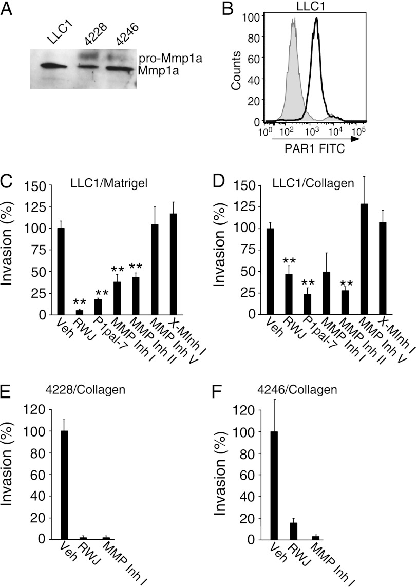
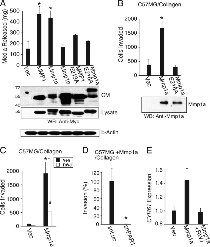
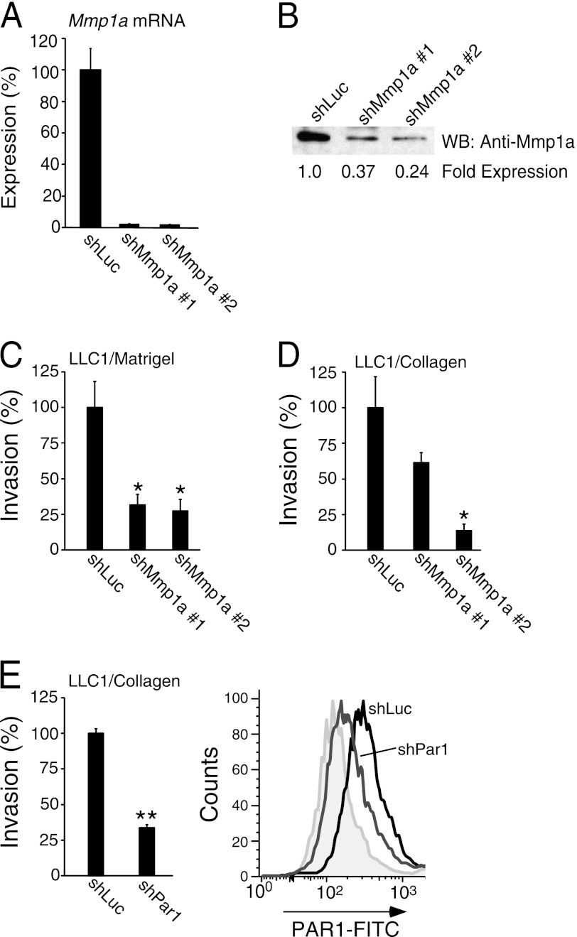
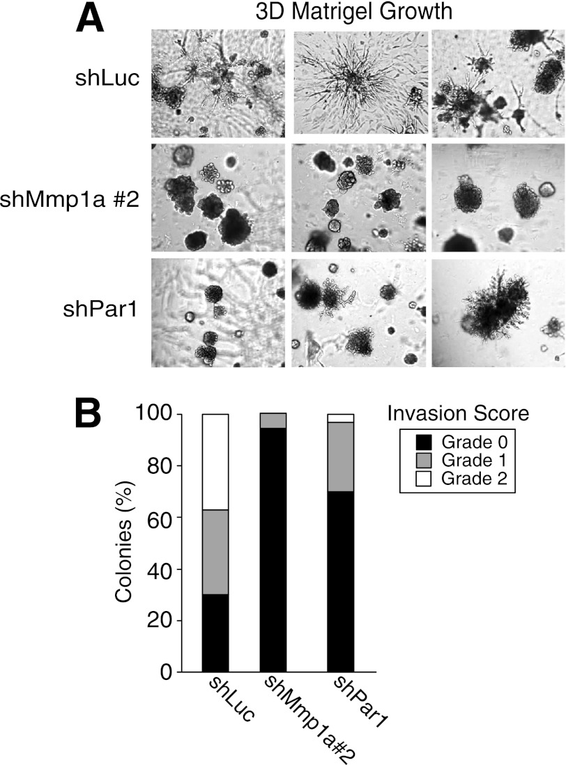
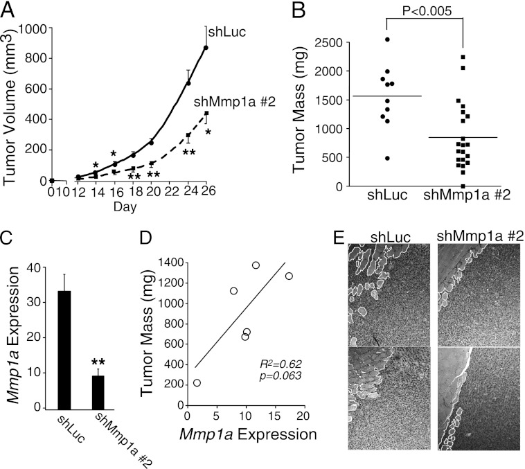
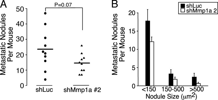
Matrix metalloprotease-1 (MMP1), a collagenase and activator of the G protein-coupled protease activated receptor-1 (PAR1), is an emerging new target implicated in oncogenesis and metastasis in diverse cancers. However, the functional mouse homologue of MMP1 in cancer models has not yet been clearly defined. We report here that Mmp1a is a functional MMP1 homologue that promotes invasion and metastatic progression of mouse lung cancer and melanoma. LLC1 (Lewis lung carcinoma) and primary mouse melanoma cells harboring active BRAF express high levels of endogenous Mmp1a, which is required for invasion through collagen. Silencing of either Mmp1a or PAR1 suppressed invasive stellate growth of lung cancer cells in three-dimensional matrices. Conversely, ectopic expression of Mmp1a conferred an invasive phenotype in epithelial cells that do not express endogenous Mmp1a. Consistent with Mmp1a acting as a PAR1 agonist in an autocrine loop, inhibition or silencing of PAR1 resulted in a loss of the Mmp1a-driven invasive phenotype. Knockdown of Mmp1a on tumor cells resulted in significantly decreased tumorigenesis, invasion, and metastasis in xenograft models. Together, these data demonstrate that cancer cell-derived Mmp1a acts as a robust functional homologue of MMP1 by conferring protumorigenic and metastatic behavior to cells.


Figures

FIGURE 1.
FIGURE 1.
Endogenous Mmp1a and PAR1 regulate invasion of mouse lung cancer and melanoma cells. A, Western blot analysis of secreted Mmp1a (pro-Mmp1a ∼ 56 kDa, Mmp1a ∼ 46 kDa) in media from LLC1 lung cancer and 4228 or 4246 melanoma cells. B, flow cytometry analysis of PAR1 surface expression on LLC1 cells using S19 FITC-PAR1 Ab versus isotype control (gray). C and D, LLC1 invasion through Matrigel (C) or type I collagen (D) in the absence or presence of PAR1 inhibitors (P1pal-7 (5 μm) or RWJ-58259 (3 μm), or MMP inhibitor (MMP Inh I) (3 μm) and MMP Inh II (5 μm), MMP Inh V (1 μm) and X-MInhI (3 μm)). All data represent mean ± S.E. of three experiments. **, p < 0.005. Veh, vehicle.
FIGURE 2.
FIGURE 2.
Mmp1a confers collagenase activity and invasive behavior through PAR1. A, collagenase activity of MMP-Myc transfected HEK293T cells plated in type I collagen gels as measured by the conversion of collagen gel to liquid. Corresponding MMP protein expression in the conditioned media (CM; 40 μl) and lysates (40 μg) was determined by Western blot (lower panel). B, invasion of Mmp1a-null C57MG cells ectopically expressing Mmp1a, inactive E216A Mmp1a, or vector control, through type I collagen toward a gradient of 10% FBS. Corresponding MMP protein expression in the conditioned media was determined by Western blot (WB; lower panel). C and D, Mmp1a-driven invasion of C57MG cells requires PAR1 activity. C, C57MG cells ectopically expressing Mmp1a or vector control, were allowed to invade through type I collagen toward a gradient of 10% FBS, in the presence or absence of the PAR1 inhibitor RWJ-58259 (5 μm). D, Mmp1a-driven invasion of C57MG cells ectopically expressing Mmp1a following stable transduction with a PAR1-targeted shRNA (shPAR1) or control (shLuc). Cells were allowed to invade through type I collagen. E, induction of a PAR1-target gene, CYR61, in Mmp1a-null C57MG cells in response to conditioned media from Mmp1a-null C57MG (vec) or C57MG cells ectopically expressing Mmp1a in the presence or absence of RWJ-58259 (5 μm). Data represent means ± S.E. of three experiments. *, p < 0.05; #, p < 0.10. Veh, vehicle.
FIGURE 3.
FIGURE 3.
Knockdown of Mmp1a-PAR1 decreases invasion of LLC1 lung cancer cells. A and B, Mmp1a mRNA (A) and Mmp1a protein expression (B) following stable, lentiviral knockdown in LLC1 cells with Mmp1a-targeted shRNA (shMmp1a-1 or -2) versus shLuc (luciferase) control. C and D, Matrigel (C) and collagen (D) invasion of Mmp1a knockdown LLC1 cells. E, LLC1 cell invasion through collagen following stable transduction with a PAR1-targeted shRNA (shPar1) versus shLuc control (left panel), with FACS analysis of PAR1 surface expression (right panel) using S19 FITC-PAR1 Ab versus isotype control (gray). All data represent means ± S.E. of three experiments. *, p < 0.05; **, p < 0.005. WB, Western blot.
FIGURE 4.
FIGURE 4.
Mmp1a and PAR1 are required for invasive stellate colony formation of lung cancer cells in three-dimensional matrices. A, growth and invasion of LLC1 cells transduced with control (shLuc), Mmp1a knockdown (shMmp1a-2), or PAR1 knockdown (shPar1) after 7 days in three-dimensional (3D) Matrigel cultures. Digital images were acquired at 60× magnification, n = 3). B, the grade of invasiveness was measured for each colony in three 6× fields and scored (grade 0, 1, 2) as described under “Experimental Procedures.”
FIGURE 5.
FIGURE 5.
Silencing of Mmp1a suppresses tumor growth and invasion of LLC1 lung cancer cells in mice. A, tumor growth following subcutaneous implantation of 200,000 shLuc (n = 10) or shMmp1a-2 (n = 21) transduced LLC1 cells in the abdominal fat pads of C57BL/6 female mice. B, mass of excised LLC1 tumors at the day 26 end point. C, Mmp1a mRNA expression in whole tumor homogenates (n = 6 per cohort) as determined by real-time PCR and expressed relative to Mmp1a mRNA levels in cultured shLuc LLC1 cells (1-fold). D, correlation between tumor size and Mmp1a mRNA expression of excised shMmp1a-2 tumors. E, invasion of shLuc or shMmp1a LLC1 transduced tumors into the abdominal musculature of mice as assessed by H&E-stained sections from subcutaneous tumors (10× magnification). Tumor/muscle interface is outlined in white. Values shown are mean ± S.E. *, p < 0.05; **, p < 0.01.
FIGURE 6.
FIGURE 6.
Silencing of Mmp1a reduces experimental metastasis of lung cancer cells in mice. A and B, number (A) and size (B) of metastatic lung nodules per mouse as determined by sum of three coronal sections per animal. LLC1 cells transduced with shMmp1a-2 or shLuc (1 × 10⁶) were injected into the tail vein of C57BL/6 mice, and lungs were harvested 28 days later and analyzed for metastases by histology. n = 10 per cohort.

References

    1. Ugalde A. P., Ordóñez G. R., Quirós P. M., Puente X. S., López-Otín C. (2010) Metalloproteases and the degradome. Methods Mol. Biol. 622, 3–29 - PubMed
    1. Rodríguez D., Morrison C. J., Overall C. M. (2010) Matrix metalloproteinases: What do they not do? New substrates and biological roles identified by murine models and proteomics. Biochim. Biophys. Acta 1803, 39–54 - PubMed
    1. Fanjul-Fernández M., Folgueras A. R., Cabrera S., López-Otín C. (2010) Matrix metalloproteinases: Evolution, gene regulation, and functional analysis in mouse models. Biochim. Biophys. Acta 1803, 3–19 - PubMed
    1. Minond D., Lauer-Fields J. L., Cudic M., Overall C. M., Pei D., Brew K., Visse R., Nagase H., Fields G. B. (2006) The roles of substrate thermal stability and P2 and P1′ subsite identity on matrix metalloproteinase triple-helical peptidase activity and collagen specificity. J. Biol. Chem. 281, 38302–38313 - PubMed
    1. Brinckerhoff C. E., Rutter J. L., Benbow U. (2000) Interstitial collagenases as markers of tumor progression. Clin. Cancer Res. 6, 4823–4830 - PubMed

Publication types