. 2012 Jun 29;287(27):22549-59.
doi: 10.1074/jbc.M111.335935. Epub 2012 May 9.

Collagen XII and XIV, new partners of cartilage oligomeric matrix protein in the skin extracellular matrix suprastructure

Affiliations

Collagen XII and XIV, new partners of cartilage oligomeric matrix protein in the skin extracellular matrix suprastructure

Pallavi Agarwal et al. J Biol Chem. .

Abstract

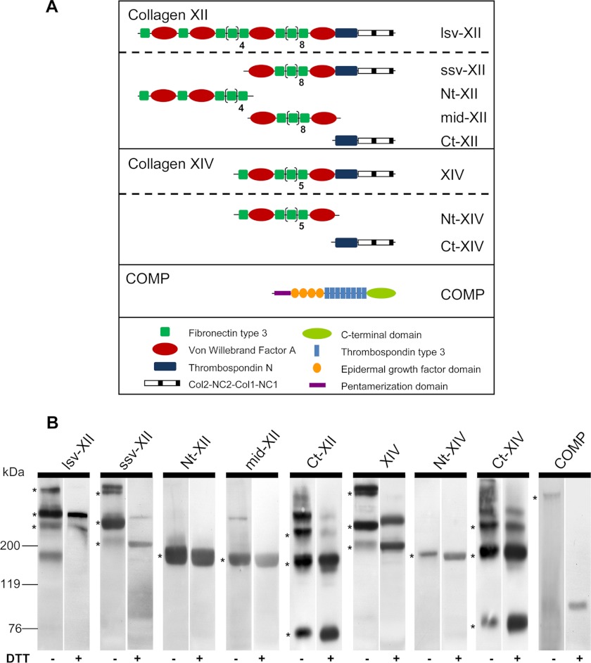
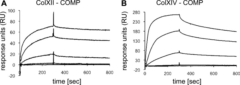
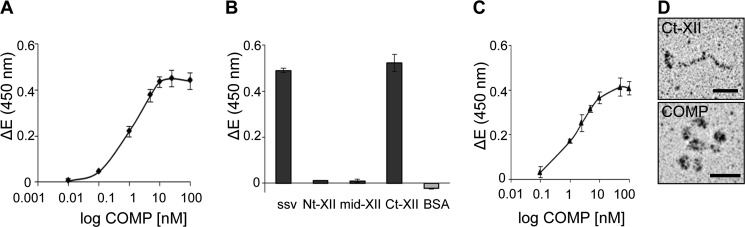
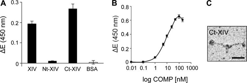
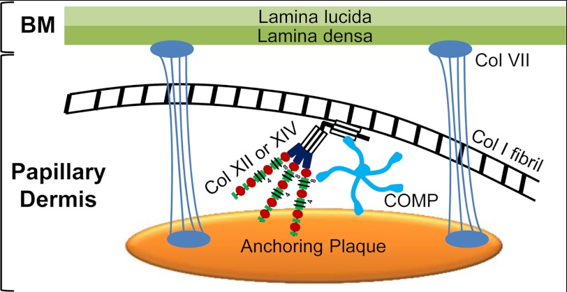
The tensile and scaffolding properties of skin rely on the complex extracellular matrix (ECM) that surrounds cells, vasculature, nerves, and adnexus structures and supports the epidermis. In the skin, collagen I fibrils are the major structural component of the dermal ECM, decorated by proteoglycans and by fibril-associated collagens with interrupted triple helices such as collagens XII and XIV. Here we show that the cartilage oligomeric matrix protein (COMP), an abundant component of cartilage ECM, is expressed in healthy human skin. COMP expression is detected in the dermal compartment of skin and in cultured fibroblasts, whereas epidermis and HaCaT cells are negative. In addition to binding collagen I, COMP binds to collagens XII and XIV via their C-terminal collagenous domains. All three proteins codistribute in a characteristic narrow zone in the superficial papillary dermis of healthy human skin. Ultrastructural analysis by immunogold labeling confirmed colocalization and further revealed the presence of COMP along with collagens XII and XIV in anchoring plaques. On the basis of these observations, we postulate that COMP functions as an adapter protein in human skin, similar to its function in cartilage ECM, by organizing collagen I fibrils into a suprastructure, mainly in the vicinity of anchoring plaques that stabilize the cohesion between the upper dermis and the basement membrane zone.


Figures

FIGURE 1.
FIGURE 1.
Domain structure and Western blot analysis of recombinant proteins. A, schematic representation of the domain structure of recombinant murine collagen XII, collagen XIV, and COMP as monomers: collagen XII long splice variant (lsv-XII), short splice variant (ssv-XII), N-terminal fragment (Nt-XII), middle fragment (mid-XII), and C-terminal fragment (Ct-XII); collagen XIV full-length (XIV), N-terminal (Nt-XIV) and C-terminal fragment (Ct-XIV); and full-length COMP (COMP). B, Western blot analysis of supernatants of stably transfected HEK293-EBNA cells was carried out by SDS-PAGE separation under non-reducing (-DTT) and reducing conditions (+DTT). Recombinant proteins were detected with antibodies recognizing the StrepII tag or the His tag (Ct fragments). COMP pentamers and trimers, dimers, and monomers of lsv-XII, ssv-XII, Ct-XII, XIV, and Ct-XIV were detected under non-reducing conditions (asterisks). Stably linked dimers of collagen XII and XIV were seen even under reducing conditions. Single specific bands were detected for middle (mid-XII) and Nt (Nt-XII, Nt-XIV) fragments. Molecular weight standards (kDa) are shown on the left.
FIGURE 2.
FIGURE 2.
COMP binds to FACIT collagens XII and XIV. Interaction of soluble lsv-XII (A) and XIV (B) with immobilized pentameric COMP was evaluated by surface plasmon resonance spectroscopy. The curves are drawn in ascending order, reflecting the concentrations of 3, 10, 30, 100, and 300 nm of the soluble analyte. The amounts of interacting analytes were monitored by measuring the variation in plasmon resonance angle over time expressed in response units (RU).
FIGURE 3.
FIGURE 3.
COMP binds to the C-terminal collagenous domain of collagen XII but not to the NC3 domain. A, binding of soluble COMP to the immobilized short splice variant of collagen XII (ssv-XII) was determined by solid phase ELISA-style assay using our antibody against mouse COMP to detect the bound ligand. The resulting saturation curve was used to calculate the apparent KD value of about 1 nm. B, recombinant fragments of collagen XII (ssv, Nt, mid, Ct) and BSA were immobilized at a concentration of 10 μg/ml, and binding of COMP (100 nm) was determined by an ELISA-style assay using the antibody against mouse COMP. BSA served as a negative control. The measurements clearly demonstrated that the binding site for COMP is located in the collagenous region of collagen XII and not in the NC3 domain. C, Ct-XII was immobilized at 10 μg/ml, and binding of pentameric COMP in concentrations ranging from 0.1 to 100 nm was analyzed. Saturable binding curves confirmed the interaction of COMP with the C-terminal collagenous domain of collagen XII. For all measurements shown in this figure, ΔE represents the measured extinction minus the blank value. Each value depicts mean ± S.D. (n = 3). D, rotary shadowing of the interacting Ct-XII fragments and pentameric COMP indicates that the recombinant proteins maintained the appropriate conformation. Scale bars = 25 nm.
FIGURE 4.
FIGURE 4.
COMP binds to the C-terminal collagenous domain of collagen XIV. A, recombinant full-length collagen XIV (XIV), collagen XIV fragments (Nt-XIV, Ct-XIV), and BSA were coated at 10 μg/ml on microtiter plates overnight at 4 °C, and binding of COMP (100 nm) was determined by ELISA-style assay using the antibody against mouse COMP. The measurements pointed to a binding site for COMP localized in the collagenous region of collagen XIV, whereas no binding was observed with the non-collagenous Nt fragment. B, saturable binding was observed for COMP to immobilized C-terminal collagenous region of collagen XIV (Ct-XIV) as determined by ELISA-style assay, confirming the presence of the binding site for COMP in the collagenous domain of collagen XIV. For all measurements shown in this figure, ΔE represents the measured extinction minus the blank value. Each value depicts the mean ± S.D. (n = 3). C, rotary shadowing of the Ct fragment of collagen XIV demonstrated that the fragment maintained an appropriate conformation. Scale bar = 25 nm.
FIGURE 5.
FIGURE 5.
COMP is produced by dermal fibroblasts and deposited in the papillary dermis of healthy human skin. A, COMP deposition and localization in human skin as detected by immunohistochemistry (n ≥ 10). Cryosections were immunostained using antibodies against human COMP showing a subepidermal localization in the superficial papillary dermis. B, the immunostaining remained restricted to the dermal part of the skin upon separation of the epidermis from the dermis by 1 m NaCl. C and D are higher-magnification images of A and B, respectively. Scale bars = 50 μm. mRNA levels were determined by real-time RT-PCR for COMP (E and I), Thy-1 (F and J), α1(I) procollagen (G and K), and keratin-14 (K14) (H and L) in epidermis (Epi) and dermis (Derm) separated after treatment with thermolysin, in cultured HaCaT cells and human primary dermal fibroblasts (Fibs). COMP expression was detected exclusively in extracts of dermis and dermal fibroblasts. Restricted expression of Thy-1 and α1(I) procollagen only in dermis and cultured fibroblasts and absence of keratin 14 (K14) expression in fibroblast cultures confirmed the dermoepidermal separation and the purity of the cell cultures. The signals obtained were normalized to S26 mRNA used as internal control. Expression levels are expressed relative to signals in epidermis and HaCaT cells. Each value depicts mean ± S.D. Results are representative of n ≥ 3 independent experiments using different donors/cell strains.
FIGURE 6.
FIGURE 6.
COMP and FACIT collagens XII and XIV show partial codistribution in the papillary dermis of human skin. Skin biopsies from healthy human donors (n ≥ 3) were coimmunostained with antibodies recognizing human collagen XII (A, green) and human COMP (B, red) raised in guinea pig and rabbit, respectively. Nuclei were stained using DAPI (blue). Coimmunofluorescence (C, merge) demonstrated extensive codistribution of COMP and collagen XII in the papillary dermis, just below the epidermis. Consecutive skin samples were coimmunostained with antibodies recognizing human collagen XIV (D, green) and human COMP (E, red) raised in guinea pig and rabbit, respectively. Collagen XIV showed deposition throughout the dermis with strong subepidermal signals, which partially codistributed with COMP (F, merge). The broken line indicates the dermoepidermal junctional zone. epi and derm depict the epidermis and dermis, respectively. Scale bar = 10 μm.
FIGURE 7.
FIGURE 7.
Ultrastructural colocalization of COMP and FACIT collagens XII and XIV in the dermis of human skin. Human skin was incubated en bloc with a rabbit antibody against mouse COMP (A), with a guinea pig antibody against human collagen XII (B), and with a guinea pig antibody against human collagen XIV (C), followed by incubation with secondary antibodies conjugated to 1 nm colloidal gold and by gold enhancement. The localization of COMP (A), collagen XII (B), and collagen XIV (C) was associated with anchoring plaques in the papillary dermis. The broken line indicates the basement membrane. Colocalization of COMP with collagen XII (D) and collagen XIV (E) at the ultrastructural level was determined by double labeling of human skin with the respective primary antibodies followed by secondary antibodies conjugated to differently sized colloidal gold particles. For colocalization of COMP with collagen XII (D) and collagen XIV (E), gold particles of 10-nm diameter were used to detect COMP, and gold particles of 6-nm diameter were used to detect either collagen XII or XIV. Colabeling of COMP/collagen XII (D) and COMP/collagen XIV (E) is indicated by arrows. D′ and E′ show magnified images of areas marked by open arrows in D and E, respectively. Scale bars = 100 nm.
FIGURE 8.
FIGURE 8.
Proposed model for the function of COMP in skin. COMP binds to collagen XII and XIV. All these proteins cluster in and around anchoring plaques. We postulate that anchoring plaques are not only amorphous patches where anchoring fibrils end but serve as docking sites for important multimeric proteins like COMP and collagens XII and XIV. Thus, anchoring plaques may connect to interstitial collagen I fibrils via COMP and FACIT collagens XII and XIV in the papillary dermis. BM, basement membrane; col VII, collagen VII that forms anchoring fibrils.

References

    1. Kadler K. E., Baldock C., Bella J., Boot-Handford R. P. (2007) Collagens at a glance. J. Cell Sci. 120, 1955–1958 - PubMed
    1. Myllyharju J., Kivirikko K. I. (2004) Collagens, modifying enzymes and their mutations in humans, flies and worms. Trends Genet. 20, 33–43 - PubMed
    1. Sorrell J. M., Caplan A. I. (2004) Fibroblast heterogeneity. More than skin deep. J. Cell Sci. 117, 667–675 - PubMed
    1. Ezura Y., Chakravarti S., Oldberg A., Chervoneva I., Birk D. E. (2000) Differential expression of lumican and fibromodulin regulate collagen fibrillogenesis in developing mouse tendons. J. Cell Biol. 151, 779–788 - PMC - PubMed
    1. Danielson K. G., Baribault H., Holmes D. F., Graham H., Kadler K. E., Iozzo R. V. (1997) Targeted disruption of decorin leads to abnormal collagen fibril morphology and skin fragility. J. Cell Biol. 136, 729–743 - PMC - PubMed

Publication types

MeSH terms