. 2012 Jun 22;287(26):22295-304.
doi: 10.1074/jbc.M111.318881. Epub 2012 May 9.

Structural tuning of the fluorescent protein iLOV for improved photostability

Affiliations

Structural tuning of the fluorescent protein iLOV for improved photostability

John M Christie et al. J Biol Chem. .

Abstract

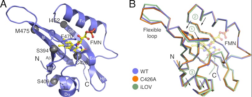
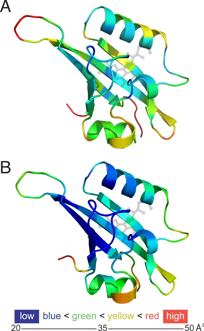
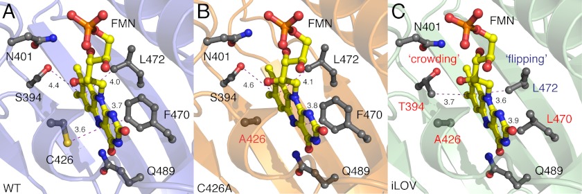
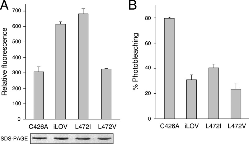
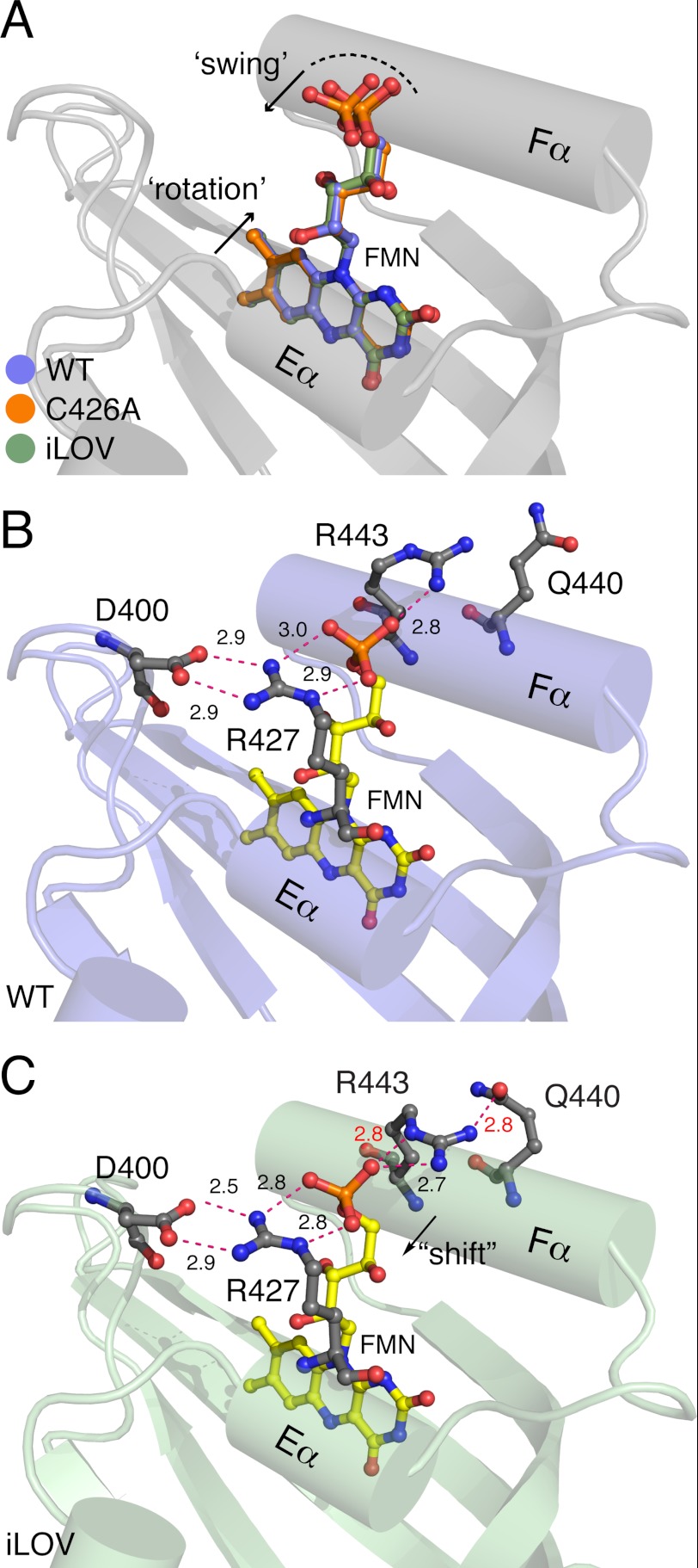
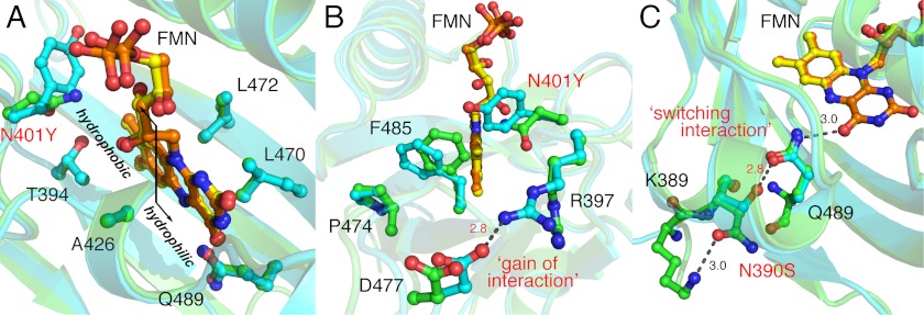
Fluorescent proteins derived from light, oxygen, or voltage (LOV) domains offer advantages over green fluorescent protein (GFP) from their small size and efficacy under anaerobic conditions. The flavoprotein improved LOV (iLOV) was engineered from the blue light receptor phototropin as a reporter of viral infection. To inform the molecular basis for the improved, photoreversible, fluorescent properties of iLOV, we employed directed evolution and determined five LOV crystallographic structures. Comparative structural analyses between iLOV and its progenitors reveal mutation-induced constraints in the environment of the flavin mononucleotide (FMN) chromophore; in iLOV, the methyl group of Thr-394 "crowds" the FMN isoalloxazine ring, Leu-470 triggers side chain "flipping" of Leu-472, and the terminal FMN phosphate shows increased anchoring. We further engineered iLOV variants that are readily detectable in bacterial and mammalian cells due to order-of-magnitude photostability increases. Structure determination of a resulting representative photostable iLOV (phiLOV) variant reveals additional constraints on the chromophore. Aromatic residues Tyr-401 and Phe-485 in phiLOV sandwich the FMN isoalloxazine ring from both sides, whereas Ser-390 anchors the side chain of FMN-interacting Gln-489 Our combined structural and mutational results reveal that constraining the FMN fluorophore yields improved photochemical properties for iLOV and its new photostable derivative. These findings provide a framework for structural fine-tuning of LOV scaffold proteins to maximize their potential as oxygen-independent fluorescent reporters.


Figures

FIGURE 1.
FIGURE 1.
Overall structures for wild-type Arabidopsis phototropin 2 LOV2 (WT), its C426A derivative, and iLOV. A, WT structure presented as ribbons (β-strands and α-helices are labeled). Residues mutated in iLOV are shown as gray spheres. FMN is presented as a yellow ball-and-stick model with red oxygen and blue nitrogen atoms. B, superposition of WT (blue), C426A (orange), and iLOV (green) structures. The carbon α trace reveals that the loop region between Aβ and Bβ (1), the β-hairpin between Hβ and Iβ (2), as well as the Cα and Dα-helices (3) are shifted in iLOV.
FIGURE 2.
FIGURE 2.
Crystallographic temperature factors (B values) mapped onto the structures of Arabidopsis phototropin 2 LOV2 (WT) (A) and iLOV (B) suggest differences in relative flexibility, both within and between these structures. Many factors can influence B values, including crystal size, x-ray exposure time, radiation dose, and crystal mosaicity, which can vary with cryo-protection protocols, yet the overall pattern of B values appears consistent with the increased packing interactions in the flavin-binding cavity, supporting decreased flexibility in iLOV relative to WT (also see supplemental Fig. S6).
FIGURE 3.
FIGURE 3.
FMN-binding cavities of wild-type Arabidopsis phototropin 2 LOV2 (WT) (A) its C426A derivative (B) and iLOV (C). Main chains are shown as ribbons. FMN is presented as a yellow ball-and-stick model with red oxygen and blue nitrogen atoms. Relevant side chains are indicated in gray as ball-and-stick models. Residues mutated in C426A and iLOV are labeled red. Critical distances between the FMN chromophore and specific residues are highlighted with dashed magenta lines. The S394T mutation in iLOV results in “crowding” of the chromophore environment at this region, whereas the side chain of Leu-472 undergoes “flipping” as a consequence of the F470L mutation.
FIGURE 4.
FIGURE 4.
Impact of Leu-472 on iLOV fluorescence. A, in vivo fluorescence of E. coli liquid cultures expressing C426A, iLOV, or iLOV harboring L472V or L472L mutations. B, fluorescence photobleaching of iLOV and respective mutants expressed in E. coli liquid culture. Fluorescence emission intensities were recorded at 495 nm upon excitation with blue light (450 nm). Values represent the mean ± S.D. (n = 3). Protein expression levels are shown in A.
FIGURE 5.
FIGURE 5.
FMN movement in iLOV when compared with wild-type Arabidopsis phototropin 2 LOV2 (WT) and its C426A derivative. Relevant side chains are indicated in gray as ball-and-stick models. Critical distances between residues and the FMN chromophore are highlighted with dashed red lines. A, superposition of the FMN of iLOV (green), WT (pale blue), and C426A (orange) reveals that the isoalloxazine ring is tilted and undergoes some “rotation” when compared with that of WT and C426A. In addition, the phosphoribityl tail of iLOV appears to “swing” and is shifted within the LOV scaffold. B and C, when compared with WT (B), the terminal phosphate of the FMN in iLOV (C) is tightly packed by two salt bridges that comprise two highly conserved arginine residues, Arg-427 and Arg-443, respectively.
FIGURE 6.
FIGURE 6.
Photobleaching of iLOV and screening for improved photostability. A, fluorescence emission spectra of iLOV in liquid cultures of E. coli before photobleaching, after bleaching, and after recovery after 5 min. Spectra were recorded by using an excitation wavelength of 450 nm. B, recovery kinetics for iLOV fluorescence in E. coli after photobleaching. Recovery fits to a first exponential and indicates a half-maximal recovery time of 75 s. C, iLOV fluorescence in E. coli colonies before photobleaching, after bleaching (dashed area), and after recovery after 5 min. D, fluorescence photobleaching of iLOV mutants isolated after one round of mutagenesis and screening for improved photostability. The dashed line represents the cut-off whereby mutant sequences exhibiting greater than 4-fold improvements in photostability were used for a second round of mutagenesis. Fluorescence emission intensities were recorded at 495 nm upon excitation with blue light (450 nm). Values represent the mean ± S.D. (n = 3).
FIGURE 7.
FIGURE 7.
Characterization of phiLOV2 variants. A, fluorescence photobleaching of variants isolated after a second round of mutagenesis and screening for improved photostability. Fluorescence emission intensities were recorded at 495 nm upon excitation with blue light (450 nm). Values represent the mean ± S.D. (n = 3). B, fluorescence of phiLOV2 variants in E. coli grown on agar medium before photobleaching and after bleaching. C, fluorescence of E. coli liquid cultures expressing phiLOV2 variants. Fluorescence emission intensities were recorded as in A. Protein expression levels are shown. D, fluorescence emission spectra of phiLOV2 variants in liquid cultures of E. coli. Spectra were recorded by using an excitation wavelength of 450 nm. E, fluorescence imaging of free iLOV and phiLOV2.1 expressed in BSC1 monkey kidney cells. F, photobleaching kinetics of iLOV and phiLOV2.1 expressed in BSC1 monkey kidney cells. Fluorescence loss in response to repeated scanning was monitored as described under “Experimental Procedures.” Values represent the mean ± S.E. (n = 2 for iLOV, n = 3 for phiLOV2.1).
FIGURE 8.
FIGURE 8.
Structural evolution of phiLOV2.1. Superposition of the FMN-binding cavity of iLOV (green) and phiLOV2.1 (pale blue) is shown. FMN is presented as a yellow ball-and-stick model for iLOV and orange for phiLOV2.1. Relevant side chains are indicated as ball-and-stick models. A, phiLOV2.1 maintains iLOV-like FMN binding properties including the flipping of Leu-472 and crowding of the immediate FMN environment by Thr-394. B, the N401Y substitution in phiLOV2.1 (labeled red) influences the chromophore binding environment at the hydrophobic side of the FMN isoalloxazine ring. Arg-397 in phiLOV2.1 is oriented to bridge the edge of the FMN-binding cleft by “gaining interaction” and hydrogen-bonding with Asp-477. C, the N390S mutation in phiLOV2.1 (labeled red) indirectly stabilizes the FMN chromophore by gaining interaction with the side chain of the FMN-interacting residue Gln-489. Critical distances between residues are highlighted with dashed lines.

References

    1. Shaner N. C., Patterson G. H., Davidson M. W. (2007) Advances in fluorescent protein technology. J. Cell Sci. 120, 4247–4260 - PubMed
    1. Tsien R. Y. (1998) The green fluorescent protein. Annu. Rev. Biochem. 67, 509–544 - PubMed
    1. Chapman S., Faulkner C., Kaiserli E., Garcia-Mata C., Savenkov E. I., Roberts A. G., Oparka K. J., Christie J. M. (2008) The photoreversible fluorescent protein iLOV outperforms GFP as a reporter of plant virus infection. Proc. Natl. Acad. Sci. U.S.A. 105, 20038–20043 - PMC - PubMed
    1. Drepper T., Eggert T., Circolone F., Heck A., Krauss U., Guterl J. K., Wendorff M., Losi A., Gärtner W., Jaeger K. E. (2007) Reporter proteins for in vivo fluorescence without oxygen. Nat. Biotechnol. 25, 443–445 - PubMed
    1. Shu X., Lev-Ram V., Deerinck T. J., Qi Y., Ramko E. B., Davidson M. W., Jin Y., Ellisman M. H., Tsien R. Y. (2011) A genetically encoded tag for correlated light and electron microscopy of intact cells, tissues, and organisms. PLoS. Biol. 9, e1001041. - PMC - PubMed

Publication types