. 2012 Jul 15;18(14):3961-3971.
doi: 10.1158/1078-0432.CCR-12-0002. Epub 2012 May 9.

Rapid angiogenesis onset after discontinuation of sunitinib treatment of renal cell carcinoma patients

Affiliations

Rapid angiogenesis onset after discontinuation of sunitinib treatment of renal cell carcinoma patients

Arjan W Griffioen et al. Clin Cancer Res. .

Abstract

Purpose: To investigate the angiogenic changes in primary tumor tissue of renal cell carcinoma (RCC) patients treated with VEGF-targeted therapy.

Experimental design: Phase II trials of VEGF pathway-targeted therapy given before cytoreductive surgery were carried out with metastatic RCC patients with the primary tumor in situ to investigate the necessity of nephrectomy. Primary tumor tissues were obtained and assessed for angiogenesis parameters. Results were compared with similar analyses on untreated tumors.

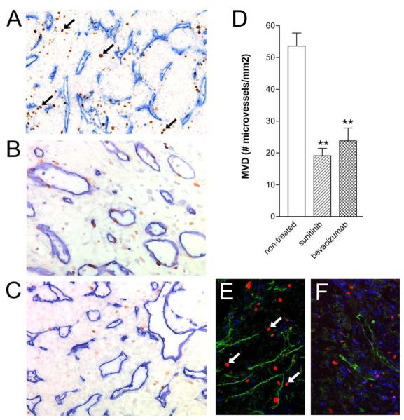
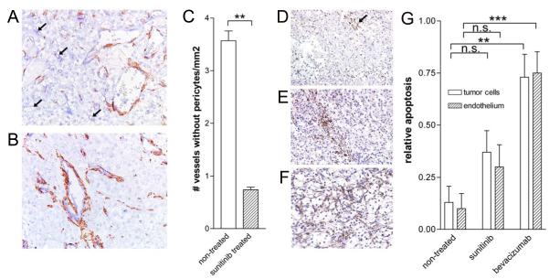
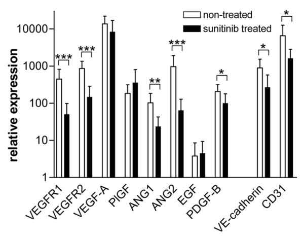
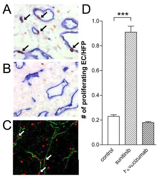
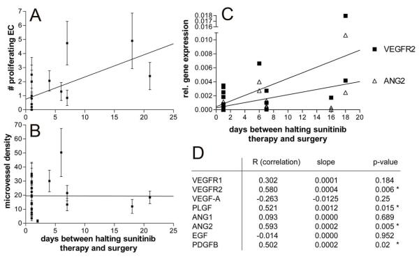
Results: Sunitinib or bevacizumab pretreatment resulted in a significant reduction of microvessel density in the primary tumor. Also, an increase in vascular pericyte coverage was found in sunitinib-pretreated tumors, consistent with efficient angiogenesis inhibition. Expression of several key regulators of angiogenesis was found to be suppressed in pretreated tissues, among which VEGFR-1 and VEGFR-2, angiopoietin-1 and angiopoietin-2 and platelet-derived growth factor-B. In addition, apoptosis in tumor and endothelial cells was induced. Interestingly, in sunitinib-pretreated tissues a dramatic increase of the number of proliferating endothelial cells was observed, which was not the case in bevacizumab-pretreated tumors. A positive correlation with the interval between halting the therapy and surgery was found, suggesting a compensatory angiogenic response caused by the discontinuation of sunitinib treatment.

Conclusion: This study describes, for the first time, the angiostatic response in human primary renal cancers at the tissue level upon treatment with VEGF-targeted therapy. Discontinuation of treatment with tyrosine kinase inhibitors leads to accelerated endothelial cell proliferation. The results of this study contribute important data to the ongoing discussion on the discontinuation of treatment with kinase inhibitors.


Figures

Figure 1
Figure 1
Suppressed microvessel density in the primary tumor after 2 cycles of sunitinib treatment. Primary tumor tissue sections of patients with RCC (n=21) were stained with a mixture of CD31/CD34 antibodies to visualize blood vessels (blue), and with anti-Ki-67 (brown, arrows) to monitor proliferation status of both the endothelial- and tumor cell compartments. Representative tissues of untreated (n=70, A), sunitinib treated (n=24, B) and bevacizumab treated (n=29, C) tissues are shown (n values are the number patient tissues with interpretable stainings). Panel D shows the quantification (means ± SEM) of all three patient groups (**p=0.001). Panels E (untreated) and F (sunitinib treated) represent immunofluorescence images, CD31/34 showing in green, Ki-67 in red, and CD3 in blue. Arrows indicate Ki-67-positive nuclei of proliferating EC.
Figure 2
Figure 2
Pericyte coverage and apoptosis is enhanced after sunitinib treatment. Panels A and B. CD31/34 (blue) and smooth muscle actin (SMA, brown) stained RCC tissues. Arrows in A indicate small blood vessels that are not associated with pericytes. C. Quantification (means ± SEM) of the number of vessels lacking pericyte coverage (identified by arrows, ** represents statistical significance, p<0.0001). D-F. PARP staining (arrow in D) of non-treated, sunitinib treated and bevacizumab treated tumors, respectively. G. Quantification of the PARP staining in tumor and endothelial cell compartments (mean ± SEM, **p<0.002, ***p<0.001).
Figure 3
Figure 3
Regulation of gene expression in the primary tumor tissue after treatment with sunitinib. Real-time quantitative PCR was performed for 10 angiogenesis related genes, i.e. VEGF receptor-1 and -2, VEGF-A, placental growth factor (PLGF), angiopoietin (ANG)-1 and -2, epithelial growth factor (EGF), platelet derive growth factor (PDGF)-B, VE-cadherin and CD31. Measurements were performed in RNA preparations of cryo-tumor tissue of sunitinib treated (n=12) and untreated (n=5) patients. Relative expression is shown (mean ± SEM) compensated for the expression of 2 house hold genes (β-actin, cyclophilin A). Statistical significance is indicated by *p<0.05, **p<0.01 and ***p<0.001.
Figure 4
Figure 4
Enhanced numbers of proliferating endothelial cells in tissues of sunitinib treated patients. CD31/34 and Ki-67 staining by standard light microscopy (blue and brown, respectively, arrows identify proliferating nuclei of endothelial cells) and immunofluorescence microscopy (green and red, respectively, arrows identify proliferating endothelial cells) in tumor tissues of sunitinib treated patients. Number of nuclei of double stained cells is quantified in C (mean ± SEM, ***p=0.0031).
Figure 5
Figure 5
Onset of angiogenesis after discontinuation of sunitinib treatment. Positive correlation (p<0.001) between the number of proliferating endothelial cells with the time-interval between treatment stop and cytoreductive surgery (A), and lack of this correlation with microvessel density (B). Panel C represents the linear regression analyses of the gene expression with the time-period between halting the sunitinib therapy and surgery. The correlation plots in C shows the most significantly correlating genes VEGFR2 and ANG2. Correlation and p-values for these and the other tested genes are shown in Table 2.

References

    1. Griffioen AW, Molema G. Angiogenesis: potentials for pharmacologic intervention in the treatment of cancer, cardiovascular diseases, and chronic inflammation. Pharmacol Rev. 2000;52:237–68. - PubMed
    1. Carmeliet P, Jain RK. Molecular mechanisms and clinical applications of angiogenesis. Nature. 2011;473:298–307. - PMC - PubMed
    1. Dings RP, Loren M, Heun H, McNiel E, Griffioen AW, Mayo KH, et al. Scheduling of radiation with angiogenesis inhibitors anginex and Avastin improves therapeutic outcome via vessel normalization. Clin Cancer Res. 2007;13:3395–402. - PMC - PubMed
    1. Dings RP, Vang KB, Castermans K, Popescu F, Zhang Y, Oude Egbrink MG, et al. Enhancement of T-cell-mediated antitumor response: angiostatic adjuvant to immunotherapy against cancer. Clin Cancer Res. 2011;17:3134–45. - PMC - PubMed
    1. Teicher BA, Holden SA, Ara G, Korbut T, Menon K. Comparison of several antiangiogenic regimens alone and with cytotoxic therapies in the Lewis lung carcinoma. Cancer Chemother Pharmacol. 1996;38:169–77. - PubMed

MeSH terms