

Computer-assisted image analysis of human cilia and Chlamydomonas flagella reveals both similarities and differences in axoneme structure
- PMID: 22573610
- PMCID: PMC3423584
- DOI: 10.1002/cm.21035
Computer-assisted image analysis of human cilia and Chlamydomonas flagella reveals both similarities and differences in axoneme structure
Abstract
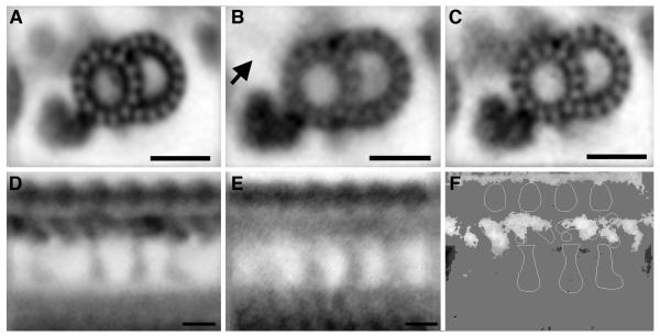
In the past decade, investigations from several different fields have revealed the critical role of cilia in human health and disease. Because of the highly conserved nature of the basic axonemal structure, many different model systems have proven useful for the study of ciliopathies, especially the unicellular, biflagellate green alga Chlamydomonas reinhardtii. Although the basic axonemal structure of cilia and flagella is highly conserved, these organelles often perform specialized functions unique to the cell or tissue in which they are found. These differences in function are likely reflected in differences in structural organization. In this work, we directly compare the structure of isolated axonemes from human cilia and Chlamydomonas flagella to identify similarities and differences that potentially play key roles in determining their functionality. Using transmission electron microscopy and 2D image averaging techniques, our analysis has confirmed the overall structural similarity between these two species, but also revealed clear differences in the structure of the outer dynein arms, the central pair projections, and the radial spokes. We also show how the application of 2D image averaging can clarify the underlying structural defects associated with primary ciliary dyskinesia (PCD). Overall, our results document the remarkable similarity between these two structures separated evolutionarily by over a billion years, while highlighting several significant differences, and demonstrate the potential of 2D image averaging to improve the diagnosis and understanding of PCD.
Copyright © 2012 Wiley Periodicals, Inc.
Figures
Similar articles
-
Comparative structural analysis of eukaryotic flagella and cilia from Chlamydomonas, Tetrahymena, and sea urchins.J Struct Biol. 2012 May;178(2):199-206. doi: 10.1016/j.jsb.2012.02.012. Epub 2012 Mar 3. J Struct Biol. 2012. PMID: 22406282
-
3D structural analysis of flagella/cilia by cryo-electron tomography.Methods Enzymol. 2013;524:305-23. doi: 10.1016/B978-0-12-397945-2.00017-2. Methods Enzymol. 2013. PMID: 23498747
-
Functional diversity of axonemal dyneins as studied in Chlamydomonas mutants.Int Rev Cytol. 2002;219:115-55. doi: 10.1016/s0074-7696(02)19012-7. Int Rev Cytol. 2002. PMID: 12211628 Review.
-
Defects in the cytoplasmic assembly of axonemal dynein arms cause morphological abnormalities and dysmotility in sperm cells leading to male infertility.PLoS Genet. 2021 Feb 26;17(2):e1009306. doi: 10.1371/journal.pgen.1009306. eCollection 2021 Feb. PLoS Genet. 2021. PMID: 33635866 Free PMC article.
-
Cilia and models for studying structure and function.Proc Am Thorac Soc. 2011 Sep;8(5):423-9. doi: 10.1513/pats.201103-027SD. Proc Am Thorac Soc. 2011. PMID: 21926393 Free PMC article. Review.
Cited by
-
Motile cilia genetics and cell biology: big results from little mice.Cell Mol Life Sci. 2021 Feb;78(3):769-797. doi: 10.1007/s00018-020-03633-5. Epub 2020 Sep 11. Cell Mol Life Sci. 2021. PMID: 32915243 Free PMC article. Review.
-
Modes of flagellar assembly in Chlamydomonas reinhardtii and Trypanosoma brucei.Elife. 2014;3:e01479. doi: 10.7554/eLife.01479. Epub 2014 Jan 21. Elife. 2014. PMID: 24448408 Free PMC article.
-
Flagellar central pair assembly in Chlamydomonas reinhardtii.Cilia. 2013 Nov 27;2(1):15. doi: 10.1186/2046-2530-2-15. Cilia. 2013. PMID: 24283352 Free PMC article.
-
The nexin-dynein regulatory complex subunit DRC1 is essential for motile cilia function in algae and humans.Nat Genet. 2013 Mar;45(3):262-8. doi: 10.1038/ng.2533. Epub 2013 Jan 27. Nat Genet. 2013. PMID: 23354437 Free PMC article.
-
Evolution of Cilia.Cold Spring Harb Perspect Biol. 2017 Jan 3;9(1):a028290. doi: 10.1101/cshperspect.a028290. Cold Spring Harb Perspect Biol. 2017. PMID: 27663773 Free PMC article. Review.
References
-
- Afzelius BA, Dallai R, Lanzavecchia S, Bellon PL. Flagellar structure in normal human spermatozoa and in spermatozoa that lack dynein arms. Tissue Cell. 1995;27(3):241–7. - PubMed
-
- Avidor-Reiss T, Maer AM, Koundakjian E, Polyanovsky A, Keil T, Subramaniam S, Zuker CS. Decoding cilia function: defining specialized genes required for compartmentalized cilia biogenesis. Cell. 2004;117(4):527–39. - PubMed
-
- Badano JL, Mitsuma N, Beales PL, Katsanis N. The Ciliopathies: An Emerging Class of Human Genetic Disorders. Annu Rev Genomics Hum Genet. 2006 - PubMed
Publication types
MeSH terms
Substances
Grants and funding
LinkOut - more resources
Full Text Sources
Miscellaneous
