Comparative Study
. 2012 May 9;32(19):6501-10.
doi: 10.1523/JNEUROSCI.5871-11.2012.

Brain state dependent postinhibitory rebound in entorhinal cortex interneurons

Affiliations
Comparative Study

Brain state dependent postinhibitory rebound in entorhinal cortex interneurons

Mohit H Adhikari et al. J Neurosci. .

Abstract

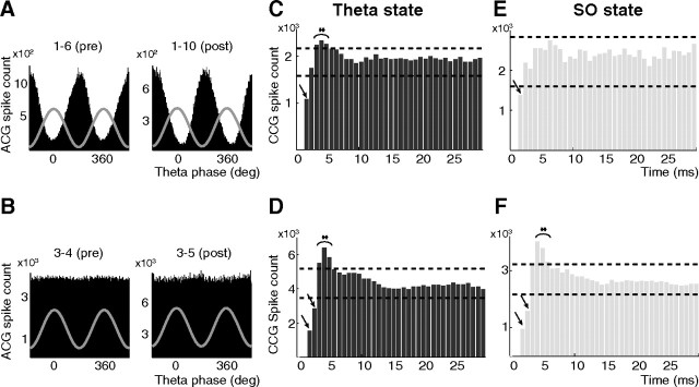
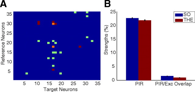
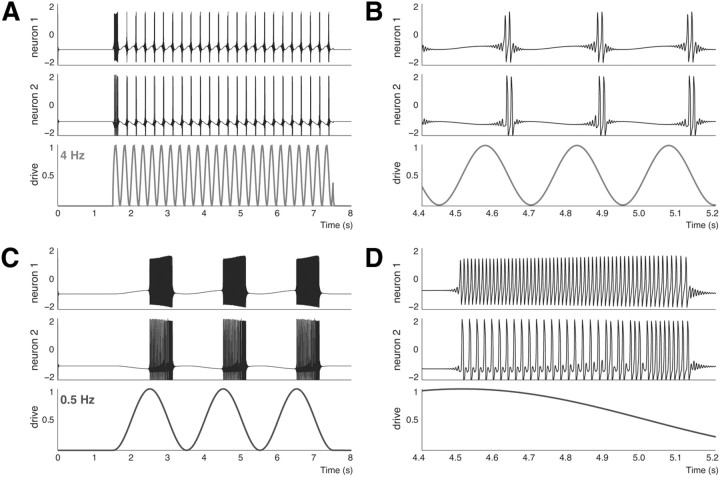
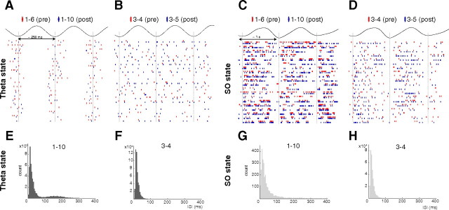
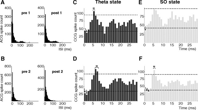
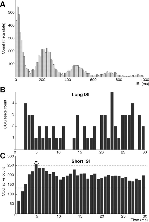
Postinhibitory rebound (PIR) is believed to play an important role in the genesis and maintenance of biological rhythms. While it has been demonstrated during several in vitro studies, in vivo evidence for PIR remains scarce. Here, we report that PIR can be observed in the dorsomedial entorhinal cortex of anesthetized rats, mostly between putatively connected GABAergic interneurons, and that it is more prevalent during the theta (4-6 Hz) oscillation state than the slow (0.5-2 Hz) oscillation state. Functional inhibition was also found to be brain state and postsynaptic cell type dependent but that alone could not explain this brain state dependence of PIR. A theoretical analysis, using two Fitzhugh-Nagumo neurons coupled to an external periodic drive, predicted that the modulation of a faster spiking rate by the slower periodic drive could account for the brain state dependence of PIR. Model predictions were verified experimentally. We conclude that PIR is cell type and brain state dependent and propose that this could impact network synchrony and rhythmogenesis.


Figures

Figure 1.
Figure 1.
Brain state dependence of PIR. A, B, Theta phase distribution of the firing of pairs of putative interneurons: neurons 1–6 (presynaptic) and 1–10 (postsynaptic) show a strong preference of their firing at the trough of the theta cycle (wave: average LFP) (A), and for neurons 3–4 and 3–5, no modulation of their firing (B). C–F, CCGs of the spike trains of the two pairs computed during the two states. Spike counts from the first two bins are not taken into account (see Materials and Methods). The arrows and braces indicate statistically significant inhibition and excitation, respectively (dashed lines, 99% global confidence intervals). Note the disappearance of PIR during SO for the 1–6/1–10 pair, while it remains significant for the 3–4/3–5 pair.
Figure 2.
Figure 2.
Verifications for the PIR hypothesis. A, Identified putative connections between all single units from one experiment. Color code: blue, no connection; red, inhibition; green, excitation; and orange, PIR. Some target neurons that display rebound upon inhibition from some interneurons (e.g., interneurons number 18 and 30 onto neuron 17) also receive excitatory connections from some principal cells (e.g., principal cells 8, 12, 15, and 21 onto neuron 17). B, Comparison between average values of the rebound strength and the extent of overlap between two sets of spikes of the target cell: those contributing to the statistically significant peaks in the PIR CCGs and those contributing to the statistically significant peaks in the CCGs for normal excitations. The low strength of overlap suggests that identified excitatory connections cannot account for the rebound excitation observed in the CCG of PIR pairs. The bar diagram considers all PIR pairs in which the target cell is also the target in at least one excitatory connection.
Figure 3.
Figure 3.
A, Responses of two neurons to an external sinusoidal drive of 4 Hz. B, Close-up on three theta cycles. Neuron 1 fires only two spikes per cycle, each of which is followed by a rebound spike of neuron 2. Thus, the number of rebound spikes is equal to the number of spikes fired by the presynaptic neuron. C, Responses of two neurons to an external sinusoidal drive of 0.5 Hz. D, Close-up on one cycle of firing activity. Note that the number of rebound spikes is less than the number of spikes fired by the presynaptic neuron. Upon closer inspection of the spikes fired by neuron 1, one can observe that the firing rate of neuron 1 is modulated by the drive. The increased firing by neuron 1 in the first part of the drive reduces the number of rebound spikes that neuron 2 can fire. Here, all the parameters are identical with the case of 4 Hz drive (i.e., I1 = 0.2, I2 = 0.0, g1 = −0.8, g2 = 0.5, g = 0.7, vth = 1.5, ksyn = 0.2).
Figure 4.
Figure 4.
Validation of model predictions. Firing patterns (A–D) of theta phase-modulated and theta phase-unmodulated cells in the data that, for sufficiently strong coupling between the drive and the neurons (here phase modulation), cells fire short and long high-frequency bursts when driven at 4 Hz (A) and 0.5 Hz (C), respectively. For weak coupling between the drive and the neurons, the cells do not show any phase modulation during the theta state (B). The most probable ISI (∼10 ms), in the ISI histograms of a theta phase-modulated interneuron (E, G) and a theta phase-unmodulated interneuron (F, H), during both states suggests a high intrinsic firing rate for both types of neurons. The ISI distribution of the theta phase-modulated interneuron during the theta state (E) is bimodal due to phase modulation.
Figure 5.
Figure 5.
ISI histograms (A) and CCGs obtained using model simulations with Gaussian noise added to each equation for sufficiently high coupling between each neuron with 4 Hz drive (C) and 0.5 Hz drive (E). The secondary peaks in the ISI histograms for the 4 Hz case reflect the theta phase modulation of firing of the neurons. PIR is statistically significant in the 4 Hz case and not in the 0.5 Hz case. ISI histograms (B) and CCGs obtained using model simulations with Gaussian noise added to each equation for sufficiently low coupling between each neuron with 4 Hz drive (D) and 0.5 Hz drive (F). The absence of secondary peaks in the ISI histograms for the 4 Hz case reflects the lack of theta phase modulation of firing of the neurons. PIR is statistically significant in both cases. Here, I1 = 0.0, I2 = 0.0, vth = −1.5, ksyn = 0.01. Mean of Gaussian noise is equal to 0, and the variance is 0.1. g1 = g2 = −1.4; g = −0.08 (A, C, E) and g1 = g2 = −1; g = −0.12 (B, D, F).
Figure 6.
Figure 6.
Rebound excitation in theta phase-modulated interneurons is not caused by the synchronization between the neuronal firing rate and the theta rhythm. A, Probability distribution of the interspike intervals of a typical theta phase-modulated interneuron during theta state. Note the peaks at the harmonic of theta frequency, which suggest possible intervals of synchronization between the neuronal firing rate and the theta frequency. B, CCGs obtained using spikes with ISI >100 ms from the spike trains of a pair of PIR displaying theta phase-modulated interneurons—very low number of counts in the CCG show that the correlated firing between isolated spikes of two cells was negligible. C, CCGs obtained using spikes with ISI <100 ms from the spike trains of the same pair of interneurons.
Figure 7.
Figure 7.
Statistical significance (SS) of PIR for all pairs as a function of the number of spikes per burst of the presynaptic cell during theta state (A) and during SO state (B). Color code: Blue, Not enough spikes for obtaining significance/no PIR connection; light blue, PIR is not SS; orange, PIR is SS; red, inhibition is SS but not rebound. There are 21 pairs during theta and 15 during SO that display PIR. For theta phase-unmodulated cells, mostly from EC5 (7 of first 9, common to both states), PIR is SS for several number of spikes per burst of the presynaptic cell, suggesting a robust connection. For theta phase-modulated cells, especially from EC2 during the theta state (18–21 in A), rebound is SS for shorter bursts, while only inhibition is SS for longer bursts.
Figure 8.
Figure 8.
The strength of functional inhibition and PIR depends upon the coupling to the rhythm. Figure shows the strength of functional inhibition (A) and rebound excitation (B), as inferred from the simulated CCGs in the case of a 4 Hz drive, as a function of two coupling strength parameters: (1) g1 (=g2), coupling between the drive and each neuron, and (2) g, the inhibitory coupling from neuron 1 to neuron 2 for a fixed noise level. As g1 (=g2) increases beyond 2, the firing of both neurons is increasingly modulated. Interestingly, we see that, for high values of g and g1 (=g2), the inhibition strength increases but the corresponding rebound strength does not necessarily increase. In fact, for a sufficiently large value of g, as the coupling with the drive increases, the rebound strength decreases, suggesting that, in this case, sufficiently high modulation of neuronal firings by the drive can suppress the number of rebound spikes fired by the postsynaptic cell. Here, I1 = 0.29, I2 = 0.0, vth = −1.5, and ksyn = 0.01. Mean of Gaussian noise is equal to 0, and the variance is 0.1.

References

    1. Aizenman CD, Linden DJ. Regulation of the rebound depolarization and spontaneous firing patterns of deep nuclear neurons in slices of rat cerebellum. J Neurophysiol. 1999;82:1697–1709. - PubMed
    1. Alviña K, Walter JT, Kohn A, Ellis-Davies G, Khodakhah K. Questioning the role of rebound firing in the cerebellum. Nat Neurosci. 2008;11:1256–1258. - PMC - PubMed
    1. Barthó P, Hirase H, Monconduit L, Zugaro M, Harris KD, Buzsáki G. Characterization of neocortical principal cells and interneurons by network interactions and extracellular features. J Neurophysiol. 2004;92:600–608. - PubMed
    1. Bertrand S, Cazalets JR. Postinhibitory rebound during locomotor-like activity in neonatal rat motoneurons in vitro. J Neurophysiol. 1998;79:342–351. - PubMed
    1. Brown SP, Hestrin S. Cell-type identity: a key to unlocking the function of neocortical circuits. Curr Opin Neurobiol. 2009;19:415–421. - PMC - PubMed

Publication types

LinkOut - more resources