Comparative Study
. 2012 May 9;32(19):6517-24.
doi: 10.1523/JNEUROSCI.0720-12.2012.

Photochemical inactivation analysis of temporal dynamics of postsynaptic native AMPA receptors in hippocampal slices

Affiliations
Comparative Study

Photochemical inactivation analysis of temporal dynamics of postsynaptic native AMPA receptors in hippocampal slices

Haruyuki Kamiya. J Neurosci. .

Abstract

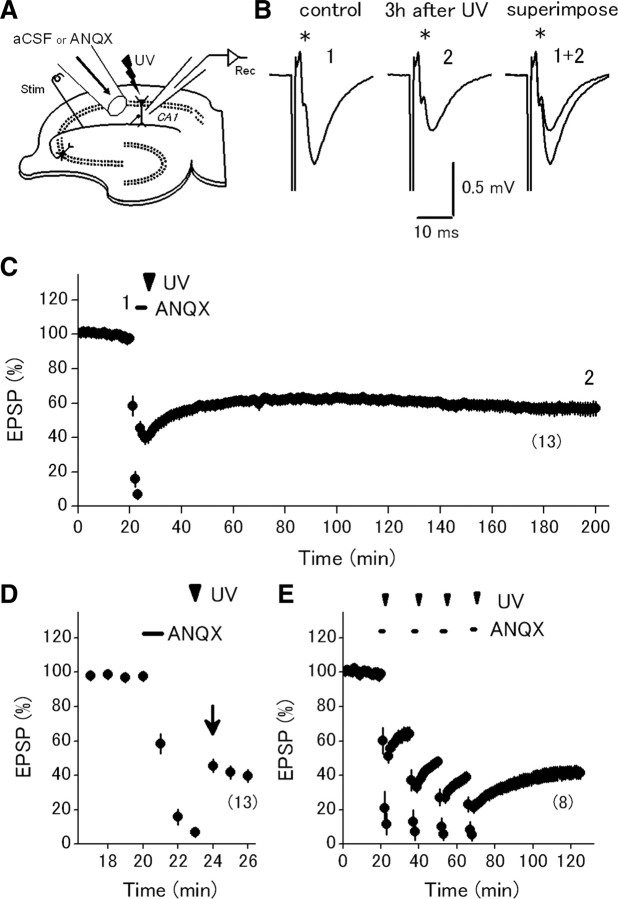
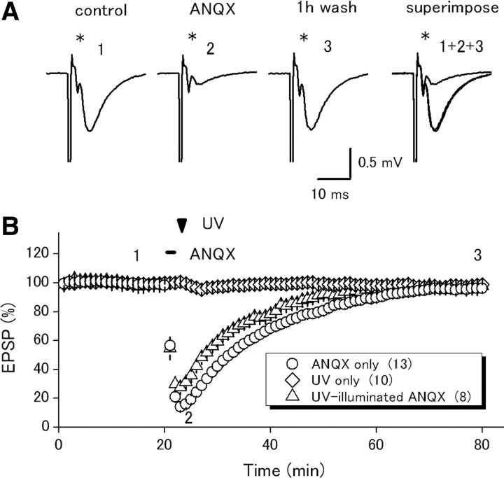
Postsynaptic expression of AMPA-type glutamate receptors (AMPAR) is more mobile than previously thought. Much evidence suggests that AMPAR are delivered from intracellular reserved pools to postsynaptic sites in a constitutive, as well as activity-dependent manner by exocytosis, lateral diffusion, or diffusional trapping. These notions were supported by optical monitoring of AMPAR subunits labeled with macromolecular tags such as GFP or Immunobeads, although it remains uncertain whether the mode and rate of synaptic delivery are similar to native "unlabeled" receptors. To reveal the real-time dynamics of native AMPAR in situ, photochemical inactivation of surface receptors using 6-azido-7-nitro-1,4-dihydroquinoxaline-2,3-dione (ANQX), a photoreactive AMPAR blocker, was adopted for acute hippocampal slices of mice. Because of the irreversible block due to cross-link formation between ANQX and surface AMPAR, recovery of EPSPs after photoinactivation reflects the time course of synaptic delivery of intracellular AMPAR. Brief UV illumination with fast application of ANQX resulted in persistent suppression of EPSPs for a prolonged period of up to 3 h, suggesting minimal synaptic delivery of AMPAR by exocytosis in the resting condition. Kinetic analysis of EPSP recovery clarified that the supply of postsynaptic AMPAR from the intracellular pool is dominated in the initial, but not in the later, phase of long-term potentiation (LTP). These results suggest that constitutive synaptic delivery is minimal in the resting condition at intact hippocampal synapses in a time scale of hours, while postsynaptic AMPAR are replaced with those in intracellular pools almost exclusively in an activity-dependent manner, typically shortly after LTP induction.


Figures

Figure 1.
Figure 1.
Photoinactivation of native AMPA receptors by photoreactive antagonist ANQX in hippocampal CA1 synapses. A, Schematic drawing of the experimental arrangement. Slice surface of the recording site is extensively perfused (at ∼0.2 ml/min) through a flow pipe with a wide open-tip diameter (250 μm). B, Effect of ANQX application with UV illumination. Specimen records show persistent suppression of EPSPs for 3 h with a minimal effect on presynaptic fiber volleys (asterisks). C, Time course of EPSPs in the above experiment. Representative traces in B are sampled at time points labeled by the numbers. D, Expanded time course around photoinactivation. EPSPs quickly increased following UV illumination (arrow), confirming that ANQX was photolyzed efficiently under these conditions. E, Effect of repeated photoinactivation 4 times at 15 min intervals. Data shown are mean ± SEM. Numbers of experiments are indicated in parentheses.
Figure 2.
Figure 2.
Reversible suppression of EPSPs by ANQX application without UV illumination. A, Effect of ANQX application alone on EPSPs at CA1 synapses. Asterisks show presynaptic fiber volleys. B, Summary of time course of EPSPs in the above experiments (open circles). Data in similar experiments with UV illumination alone are shown in the same graph (open diamonds) for reference. In another set of experiments, already UV-illuminated ANQX was applied (open triangles) to examine the effect of photoproduct FQX. Numbers of experiments are indicated in parentheses.
Figure 3.
Figure 3.
Photoinactivation of native AMPA receptors in mossy fiber-CA3 synapses. A, Schematic diagram of the experimental arrangement. Slice surface of the recording site (stratum lucidum) is locally perfused with the same flow pipe. B, Effect of ANQX application with UV illumination. Specimen records show persistent suppression of EPSPs lasting for 3 h, with a minimal effect on presynaptic fiber volleys (asterisks). C, Time course of EPSPs in the above experiment (closed circles). For comparison, data in similar experiments with ANQX application alone (open circles) or with UV illumination alone (open diamonds) are superimposed. DCG-IV (1 μm) was applied at the end of experiments to confirm that the mossy fibers were selectively stimulated. Numbers of experiments are indicated in parentheses.
Figure 4.
Figure 4.
Accelerated synaptic delivery in the initial, but not in the later, phase of LTP induced by TBS. A, Effect of photoinactivation either 5 min (open circles) or 60 min (closed circles) after TBS (10 bursts of 4 pulses at 100 Hz with an interval of 200 ms). Representative traces show data in the 60 min group. B, Expanded time course around photoinactivation applied 60 min after TBS. Data points from 27 to 77 min are curtailed for clarity. Red line represents single-exponential fit of the time course of recovery after photoinactivation. C, Same as B except that photoinactivation was applied 5 min after TBS. Numbers of experiments are indicated in parentheses.
Figure 5.
Figure 5.
LTP induction by TBS is not affected by the preceding photoinactivation. A, Effect of TBS at 60 min after photoinactivation. B, Data in A are normalized with those during the 20 min before TBS. Numbers of experiments are indicated in parentheses.
Figure 6.
Figure 6.
LTP induced by high-frequency stimulation (HFS) accelerates recovery after photoinactivation in the initial, but not in the later, phase. A, Effect of photoinactivation either 5 min (open circles) or 60 min (closed circles) after HFS (100 Hz for 1 s, three times, 20 s interval). B, Expanded time course around photoinactivation applied 60 min after HFS. Red line represents single-exponential fit of recovery. C, Same as B except that photoinactivation was applied 5 min after HFS. Numbers of experiments are indicated in parentheses.
Figure 7.
Figure 7.
Two pathway experiments for comparison of the recovery phase in potentiated and nonpotentiated inputs. A, Effect of photoinactivation on either 5 min after TBS (open circles) or nontetanized input (closed circles) in the same experiments. B, Expanded time course around photoinactivation in nontetanized input. Red line represents single-exponential fit of time course of recovery after photoinactivation. C, Same as B except that photoinactivation was applied 5 min after TBS. Numbers of experiments are indicated in parentheses.

References

    1. Adesnik H, Nicoll RA. Conservation of glutamate receptor 2-containing AMPA receptors during long-term potentiation. J Neurosci. 2007;27:4598–4602. - PMC - PubMed
    1. Adesnik H, Nicoll RA, England PM. Photoinactivation of native AMPA receptors reveals their real-time trafficking. Neuron. 2005;48:977–985. - PubMed
    1. Andrásfalvy BK, Magee JC. Changes in AMPA receptor currents following LTP induction on rat CA1 pyramidal neurones. J Physiol. 2004;559:543–554. - PMC - PubMed
    1. Ashby MC, Maier SR, Nishimune A, Henley JM. Lateral diffusion drives constitutive exchange of AMPA receptors at dendritic spines and is regulated by spine morphology. J Neurosci. 2006;26:7046–7055. - PMC - PubMed
    1. Bayazitov IT, Richardson RJ, Fricke RG, Zakharenko SS. Slow presynaptic and fast postsynaptic components of compound long-term potentiation. J Neurosci. 2007;27:11510–11521. - PMC - PubMed

Publication types

MeSH terms