Comparative Study
. 2012 May 9;32(19):6561-9.
doi: 10.1523/JNEUROSCI.3353-11.2012.

Transglutaminase inhibition protects against oxidative stress-induced neuronal death downstream of pathological ERK activation

Affiliations
Comparative Study

Transglutaminase inhibition protects against oxidative stress-induced neuronal death downstream of pathological ERK activation

Manuela Basso et al. J Neurosci. .

Erratum in

  • J Neurosci. 2012 Aug 8;32(32):11157

Abstract

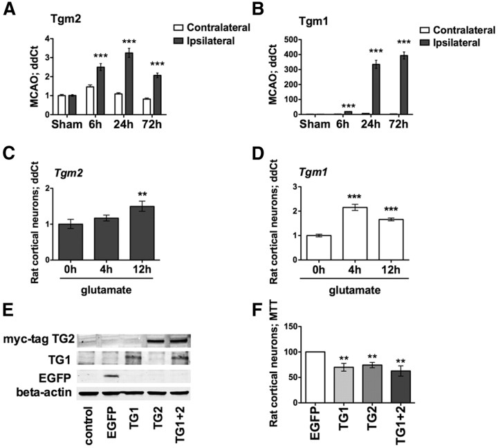
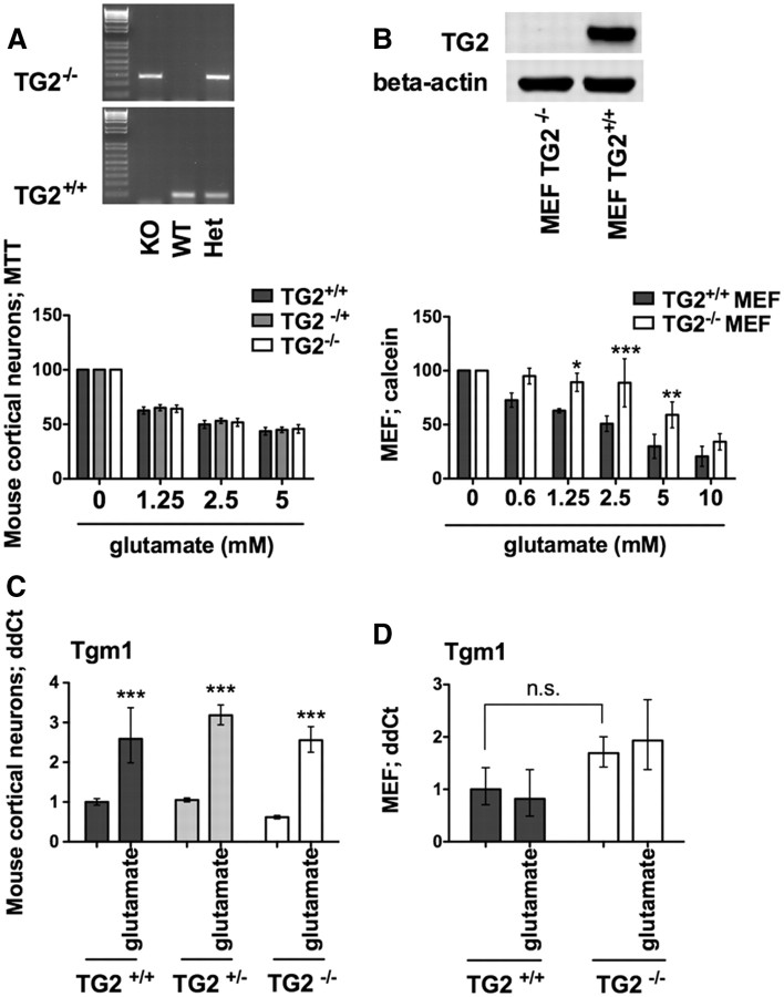
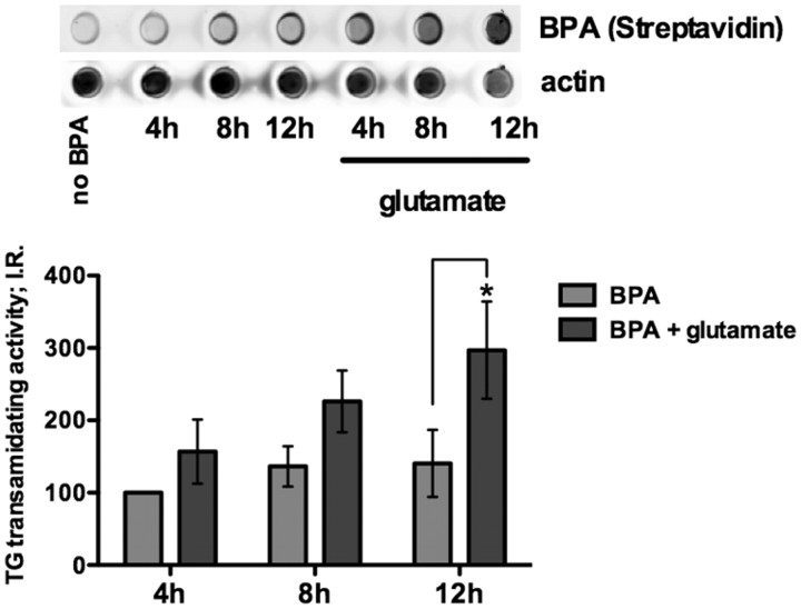
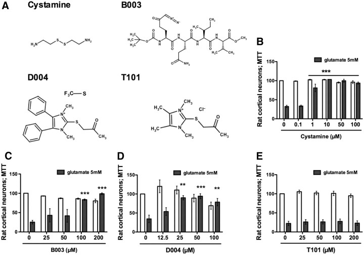
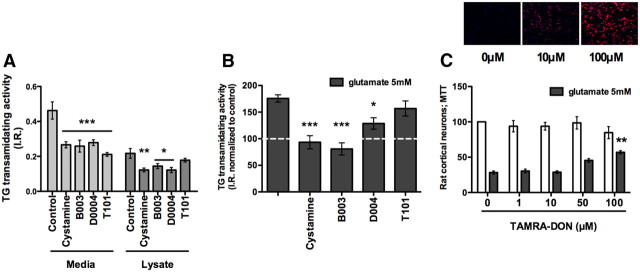
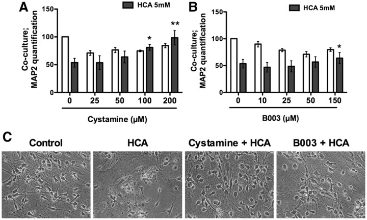
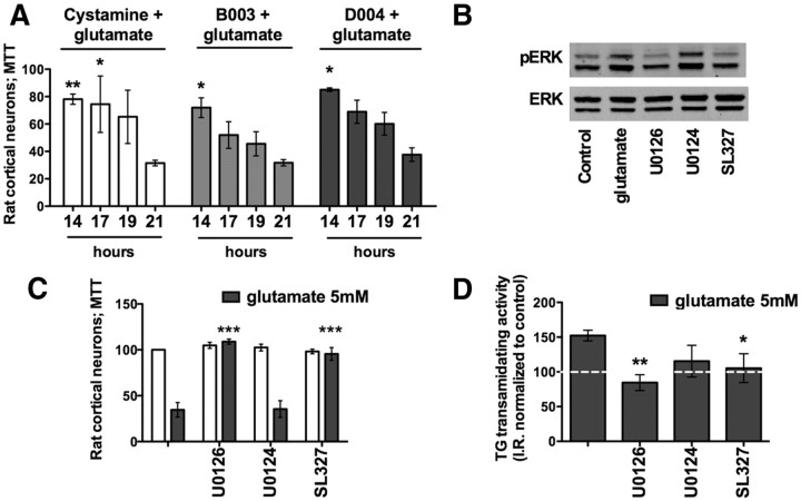
Molecular deletion of transglutaminase 2 (TG2) has been shown to improve function and survival in a host of neurological conditions including stroke, Huntington's disease, and Parkinson's disease. However, unifying schemes by which these cross-linking or polyaminating enzymes participate broadly in neuronal death have yet to be presented. Unexpectedly, we found that in addition to TG2, TG1 gene expression level is significantly induced following stroke in vivo or due to oxidative stress in vitro. Forced expression of TG1 or TG2 proteins is sufficient to induce neuronal death in Rattus norvegicus cortical neurons in vitro. Accordingly, molecular deletion of TG2 alone is insufficient to protect Mus musculus neurons from oxidative death. By contrast, structurally diverse inhibitors used at concentrations that inhibit TG1 and TG2 simultaneously are neuroprotective. These small molecules inhibit increases in neuronal transamidating activity induced by oxidative stress; they also protect neurons downstream of pathological ERK activation when added well after the onset of the death stimulus. Together, these studies suggest that multiple TG isoforms, not only TG2, participate in oxidative stress-induced cell death signaling; and that isoform nonselective inhibitors of TG will be most efficacious in combating oxidative death in neurological disorders.


Figures

Figure 1.
Figure 1.
TG1 and TG2 mRNA levels are upregulated in a focal model of stroke (MCAO) and in an in vitro model of neuronal oxidative stress, and they are sufficient to induce cell death. A, B, Temporal expression profile for Tgm2 (A) and Tgm1 (B) in MCAO. Both of the genes are significantly upregulated in the ipsilateral side compared with the contralateral hemisphere (***p < 0.001). A similar upregulation is revealed in an in vitro model of oxidative stress. C, D, Tgm2 (C) and Tgm1 (D) levels are significantly upregulated 12 h after glutamate (5 mm) exposure in rat neurons (**p < 0.01; ***p < 0.0001 compared with the relative glutamate 0 h). E, F, Forced expression of TG1 and/or TG2 (E) exerts neuronal toxicity, as revealed by MTT (F). Significant toxicity compared with EGFP expression. **p < 0.01. EGFP-expressing cells are calculated at 100% survival.
Figure 2.
Figure 2.
TG2 is necessary for oxidative death in MEFs but not cortical neurons. Resistance of cortical neurons to TG2 knockout is associated with compensatory upregulation of Tgm1. A, B, TG2−/− in single embryonic neuronal cultures (A) and in MEFs (B) and relative MTT assay. TG2 deletion in MEFs significantly protects against glutamate-induced death. *p < 0.05; **p < 0.01; ***p < 0.001 compared with TG2+/+ MEFs; untreated controls are calculated at 100% of survival. C, D, Tgm1 levels are induced after 12 h of glutamate treatment in mouse neurons (C) (***p < 0.001 compared with controls), but not in MEFs (D).
Figure 3.
Figure 3.
Intracellular TG transamidating activity is increased upon glutamate treatment. Endogenous transamidating activity is significantly increased after 12 h of glutamate treatment compared with control by dot blot assay. **p < 0.01.
Figure 4.
Figure 4.
TG inhibition protects immature cortical neurons from oxidative stress-mediated cell death. A, Structures of four diverse, reversible or irreversible, isoform-nonselective TG inhibitors tested in an in vitro model of oxidative stress. B–D, Cystamine (1–100 μm) (B), B003 (100, 200 μm) (C), and D004 (25–100 μm) (D) protect primary immature cortical neurons against oxidative stress-induced cell death. E, T101, a cell-impermeable TG inhibitor, fails to provide protection. **p < 0.01; ***p < 0.001 compared with glutamate treatment alone; untreated controls are calculated at 100% of survival.
Figure 5.
Figure 5.
Inhibition of intracellular TG activity correlates with neuroprotection. A, All inhibitors tested are able to significantly reduce extracellular transamidating activity (A). A, B, T101 fails to inhibit intracellular TG transamidating activity. B, All the protective inhibitors are able to significantly downregulate TG2 activity to control levels. C, TAMRA-DON, a peptide-based, fluorescent inhibitor analog to B003, enters the cells (10–100 μm) (top), and it protects against oxidative stress (100 μm) (bottom). **p < 0.01 compared with glutamate treatment alone; untreated controls are calculated at 100% of survival.
Figure 6.
Figure 6.
TG inhibition in astrocytes non-cell autonomously protects neurons. A, B, Astrocytes are treated overnight with the TG inhibitors Cystamine (A) or B003 (B). Following washoff of the inhibitors, neurons were plated with or without the glutamate analog HCA (5 mm) for 48 h: *p < 0.05; **p < 0.01 neuronal survival compared with glutamate treatment alone, untreated controls are calculated at 100% of survival. C, Representative phase contrast pictures for A and B; cystamine, 200 μm; B003, 150 μm.
Figure 7.
Figure 7.
TG transamidating activity is a downstream target of the ERK pathway. A, TG inhibitors exerted protection when added up to 14–17 h post-glutamate exposure. B–D, Glutamate induces phosphorylated ERK (pERK) (B); MEK inhibitors U0126 and SL327 block pERK activation (B) and protect against glutamate toxicity (C) by reducing TG activation to the control level (D). *p < 0.05; **p < 0.01 compared with glutamate treatment alone; untreated controls are calculated at 100% of survival. U0124 is a negative control for U0126.

References

    1. Akimov SS, Belkin AM. Opposing roles of Ras/Raf oncogenes and the MEK1/ERK signaling module in regulation of expression and adhesive function of surface transglutaminase. J Biol Chem. 2003;278:35609–35619. - PubMed
    1. Antonyak MA, Li B, Boroughs LK, Johnson JL, Druso JE, Bryant KL, Holowka DA, Cerione RA. Cancer cell-derived microvesicles induce transformation by transferring tissue transglutaminase and fibronectin to recipient cells. Proc Natl Acad Sci U S A. 2011;108:4852–4857. - PMC - PubMed
    1. Bailey CD, Johnson GV. The protective effects of cystamine in the R6/2 Huntington's disease mouse involve mechanisms other than the inhibition of tissue transglutaminase. Neurobiol Aging. 2006;27:871–879. - PubMed
    1. Belkin AM. Extracellular TG2: emerging functions and regulation. FEBS J. 2011;278:4704–4716. - PMC - PubMed
    1. Borrell-Pagès M, Canals JM, Cordelières FP, Parker JA, Pineda JR, Grange G, Bryson EA, Guillermier M, Hirsch E, Hantraye P, Cheetham ME, Néri C, Alberch J, Brouillet E, Saudou F, Humbert S. Cystamine and cysteamine increase brain levels of BDNF in Huntington disease via HSJ1b and transglutaminase. J Clin Invest. 2006;116:1410–1424. - PMC - PubMed

Publication types

MeSH terms

Substances