Comparative Study
. 2012 May 9;32(19):6570-7.
doi: 10.1523/JNEUROSCI.6274-11.2012.

In vivo proliferative regeneration of balance hair cells in newborn mice

Affiliations
Comparative Study

In vivo proliferative regeneration of balance hair cells in newborn mice

Joseph C Burns et al. J Neurosci. .

Abstract

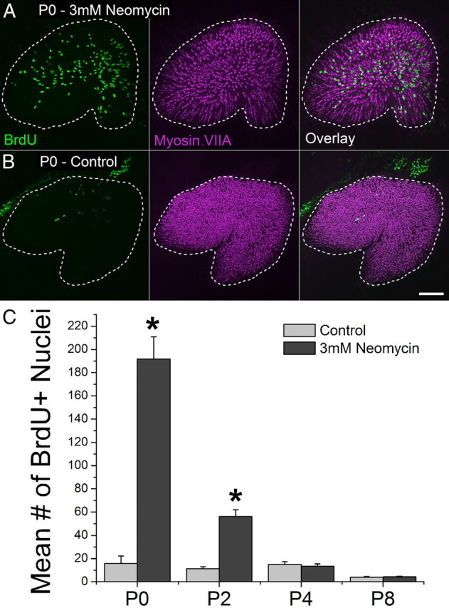
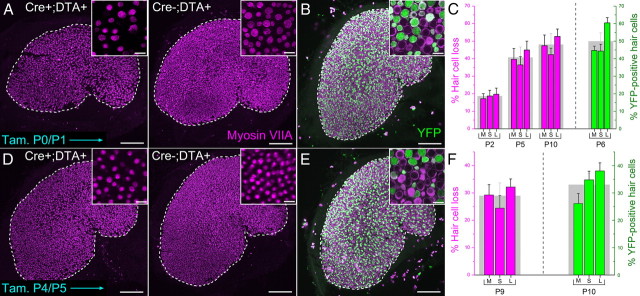
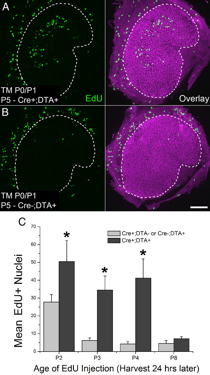
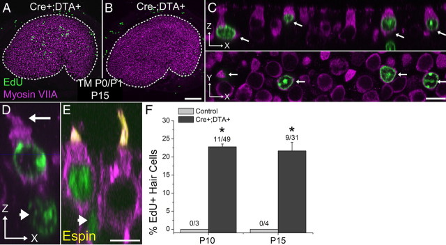
The regeneration of mechanoreceptive hair cells occurs throughout life in non-mammalian vertebrates and allows them to recover from hearing and balance deficits that affect humans and other mammals permanently. The irreversibility of comparable deficits in mammals remains unexplained, but often has been attributed to steep embryonic declines in cellular production. However, recent results suggest that gravity-sensing hair cells in murine utricles may increase in number during neonatal development, raising the possibility that young mice might retain sufficient cellular plasticity for mitotic hair cell regeneration. To test for this we used neomycin to kill hair cells in utricles cultured from mice of different ages and found that proliferation increased tenfold in damaged utricles from the youngest neonates. To kill hair cells in vivo, we generated a novel mouse model that uses an inducible, hair cell-specific CreER allele to drive expression of diphtheria toxin fragment A (DTA). In newborns, induction of DTA expression killed hair cells and resulted in significant, mitotic hair cell replacement in vivo, which occurred days after the normal cessation of developmental mitoses that produce hair cells. DTA expression induced in 5-d-old mice also caused hair cell loss, but no longer evoked mitotic hair cell replacement. These findings show that regeneration limits arise in vivo during the postnatal period when the mammalian balance epithelium's supporting cells differentiate unique cytological characteristics and lose plasticity, and they support the notion that the differentiation of those cells may directly inhibit regeneration or eliminate an essential, but as yet unidentified pool of stem cells.


Figures

Figure 1.
Figure 1.
Cells in the utricles of newborn mice respond to hair cell death with robust proliferation in vitro. A, B, Confocal images of utricles from P0 mice that were treated with 3 mm neomycin or vehicle control for 24 h and cultured for 72 h thereafter (BrdU, green, was included in the media throughout). Hair cells are labeled with an antibody to myosin VIIA (purple). A dashed line marks the macula's edge. Scale bar, 100 μm. C, Graph of BrdU-positive nuclei in the macula of neomycin-damaged and control utricles from mice of different ages, n = 8 per age. Asterisks indicate a significant difference between treatment and controls (p < 0.05).
Figure 2.
Figure 2.
Hair cells in neonates can be ablated in vivo using an inducible mouse model. A, Confocal images of utricles from an Atoh1-CreER™; ROSA26DTA/+ mouse and a ROSA26DTA/+ control mouse injected with tamoxifen at P0 and P1 and killed at P10. Hair cells are labeled with antibodies to myosin VIIA (purple). Scale bar, 100 μm. Insets show zoomed regions of the sensory epithelium. Scale bar, 10 μm. B, Confocal image of a utricle from an Atoh1-CreER™; ROSA26eYFP/+ mouse induced with tamoxifen at P0/P1 and killed at P6. eYFP is shown in green. Scale bar, 100 μm. Inset shows zoomed region of the sensory epithelium. Scale bar, 10 μm. C, Graph of the percentage difference in the density of hair cells between utricles from Atoh1-CreER™; ROSA26DTA/+ mice and control mice that were induced with tamoxifen at P0/P1 and killed at the ages indicated (bars in purple), n = 4. M, medial; S, striolar; L, lateral regions of the utricle, respectively. The gray bar behind the colored columns shows the average for the entire utricle. Also shown is the percentage of eYFP-positive hair cells in utricles from Atoh1-CreER™; ROSA26eYFP/+ mice induced with tamoxifen at P0/P1 and killed at P6 (bars in green), n = 4. Data expressed as mean ± SEM. D–F, Same as in A–C, except that mice were induced with tamoxifen at P4/P5. Utricles in D and E were fixed from mice killed at P9 and P10, respectively. Data represented by green bars in F were taken from utricles fixed at P10. Note different scales for graphs in C and F.
Figure 3.
Figure 3.
Cells in the utricles of newborn mice respond to hair cell death with robust proliferation in vivo. A, B, Confocal images of utricles from an Atoh1-CreER™; ROSA26DTA/+ mouse and a ROSA26DTA/+ control mouse induced with tamoxifen (TM) at P0/P1. EdU (green) was injected once at P4 and utricles were harvested 24 h later. Hair cells are labeled with an antibody to myosin VIIA (purple). Scale bar, 100 μm. C, Graph of the number of EdU-positive nuclei in maculae of Atoh1-CreER™; ROSA26DTA/+ or control mice of different ages that were induced with tamoxifen at P0/P1 and received EdU 24 h before utricle fixation, n = 4 per age. Data expressed as mean ± SEM. Asterisks indicate significant difference between treatment and controls (p < 0.05).
Figure 4.
Figure 4.
Damage-induced proliferation gives rise to new hair cells in vivo. A, B, Confocal image of a utricle from a P15 Atoh1-CreER™; ROSA26DTA/+ mouse and a control mouse lacking Cre that received tamoxifen (TM) at P0/P1 and EdU (green) at P4. Hair cells are labeled with an antibody to myosin VIIA (purple). Scale bar, 100 μm. C, Confocal images of a zoomed region in A. Top (x-z view), Orthogonal view along the long axis of the hair cells. Arrows indicate four EdU-positive/myosin VIIA-positive hair cells. Bottom (x-y view), Cross-sectional view taken at the level of the hair cell nuclei. Scale bar, 10 μm. D, E, High-resolution confocal images of regenerated hair cells in utricles from P15 Atoh1-CreER™; ROSA26DTA/+ mice that received tamoxifen at P0/P1 and EdU at P4. Image is parallel to the long axis of the hair cells (x-z view). Arrow marks a hair bundle, and arrowheads mark EdU-positive hair cell/supporting cell pairs. Regenerated hair cells have shorter hair bundles than preexisting hair cells, as shown by labeling with an espin antibody (D). Scale bar, 10 μm. F, Graph of the percentage of EdU-positive cells that were also positive for myosin VIIA in utricles from P10 and P15 Atoh1-CreER™; ROSA26DTA/+ or control mice that were injected once with EdU at P4 (tamoxifen given at P0/P1), n = 4. In the fractions above the bars, the mean number of EdU-positive/myosin VIIA-positive cells is shown in the numerator, and the mean number of EdU-positive cells is shown in the denominator. Data in graphs are expressed as mean ± SEM. Asterisks indicate significant difference between treatment and controls (p < 0.05).

References

    1. Abrahamsen B, Zhao J, Asante CO, Cendan CM, Marsh S, Martinez-Barbera JP, Nassar MA, Dickenson AH, Wood JN. The cell and molecular basis of mechanical, cold, and inflammatory pain. Science. 2008;321:702–705. - PubMed
    1. Baird RA, Steyger PS, Schuff NR. Mitotic and nonmitotic hair cell regeneration in the bullfrog vestibular otolith organs. Ann N Y Acad Sci. 1996;781:59–70. - PubMed
    1. Brigande JV, Heller S. Quo vadis, hair cell regeneration? Nat Neurosci. 2009;12:679–685. - PMC - PubMed
    1. Burns JC, Christophel JJ, Collado MS, Magnus C, Carfrae M, Corwin JT. Reinforcement of cell junctions correlates with the absence of hair cell regeneration in mammals and its occurrence in birds. J Comp Neurol. 2008;511:396–414. - PMC - PubMed
    1. Chen P, Segil N. p27(Kip1) links cell proliferation to morphogenesis in the developing organ of Corti. Development. 1999;126:1581–1590. - PubMed

Publication types