

doi: 10.1128/JVI.00401-12.
Epub 2012 May 9.
Infectious clones of novel lineage 1 and lineage 2 West Nile virus strains WNV-TX02 and WNV-Madagascar
Affiliations
- PMID: 22573862
- PMCID: PMC3416286
- DOI: 10.1128/JVI.00401-12
Item in Clipboard
Infectious clones of novel lineage 1 and lineage 2 West Nile virus strains WNV-TX02 and WNV-Madagascar
J Virol.
2012 Jul.
Abstract
We report the generation of West Nile virus (WNV) infectious clones for the pathogenic lineage 1 Texas-HC2002 and nonpathogenic lineage 2 Madagascar-AnMg798 strains. The infectious clones exhibited biological properties similar to those of the parental virus isolates. We generated chimeric viruses and found that viral factors within the structural and nonstructural regions of WNV-TX contribute to the control of type I interferon defenses. These infectious clones provide new reagents to study flavivirus immune regulation and pathogenesis.
Figures

The WNV-TX infectious clone (i.c.) and the WNV-MAD infectious clone display biological properties comparable to those of the parental virus isolates. BHK-21 cells were infected in triplicate at an MOI of 5.0 (A and B) or 0.01 (C and D) with parental WNV-TX (working stocks were derived from single-round plaque purification in Vero E6 cells and two rounds of amplification in HEK 293 cells) and the WNV-TX infectious clone or parental WNV-MAD (working stocks were derived from two rounds of amplification in Vero E6 cells) and the WNV-MAD infectious clone. Volumes of 100 μl of cell culture supernatant were removed at the indicated times postinfection and replaced with fresh cell culture medium. Viral burdens in the culture supernatants were then determined by plaque assay on BHK-21 cells. (E and F) Particle-to-PFU ratios were determined by triplicate viral RNA extraction from working viral stocks (harvested at 48 h postinfection) using a QIAamp viral RNA extraction kit, followed by quantitative reverse transcription-PCR with virus-specific primers. Viral RNA copy numbers were divided by the viral titers determined on BHK-21 cells. Data are representative of three independent experiments. *, P < 0.05.

The WNV-TX infectious clone (i.c.) and the WNV-MAD infectious clone exhibit replication fitness comparable to that of their respective parental virus isolates in IFN-competent cells. A549 cells were infected in triplicate at an MOI of 5.0 (A and B) or 0.01 (C and D) with the parental WNV-TX and WNV-TX infectious clones or parental WNV-MAD and WNV-MAD infectious clone. Volumes of 100 μl of cell culture supernatant were removed at the indicated times postinfection and replaced with fresh cell culture medium. Viral burdens in the collected culture supernatants were then determined by plaque assay on Vero E6 cells. (D and E) A549 cells were mock infected or infected (MOI of 5) in triplicate with the parental WNV-TX and WNV-TX infectious clones or the parental WNV-MAD and WNV-MAD infectious clones. At 6, 12, 24, or 48 h postinfection, cells were pulse treated with 1,000 IU of IFN-α (PBL IFN) for 30 min and whole-cell lysates were collected in modified radioimmunoprecipitation assay buffer (10 mM Tris [pH 7.5]–150 mM NaCl–0.5% sodium deoxycholate–1% Triton X-100 supplemented with protease inhibitor cocktail [Sigma] and phosphatase inhibitor cocktail II [Calbiochem]) and analyzed by immunoblotting to detect STAT1 phosphotyrosine residue 701 (Cell Signaling), total STAT1 (Cell Signaling), STAT2 phosphotyrosine residue 689 (Upstate), total STAT2 (Cell Signaling), WNV NS3 (R&D Systems), and glyceraldehyde 3-phosphate dehydrogenase (GAPDH; Santa Cruz). Data are representative of three independent experiments. *, P < 0.05.
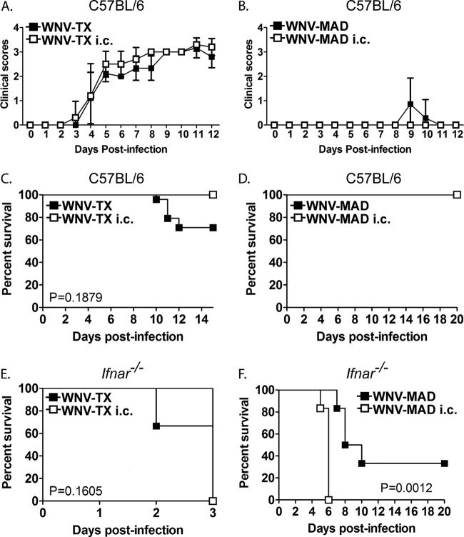
Infectious cloned viruses display in vivo biological properties comparable to those of the parental virus isolates. Six- to 12-week-old C57BL/6 mice were infected with parental WNV-TX (n = 24) and the WNV-TX infectious clone (i.c.) (n = 10) (A) or parental WNV-MAD (n = 6) and the WNV-MAD infectious clone (n = 6) (B) by subcutaneous injection into the left rear footpad of 100 PFU in a 10-μl inoculum diluted in phosphate-buffered saline supplemented with 1% heat-inactivated fetal bovine serum. Mice were monitored daily and scored for clinical signs as follows: 1, ruffled fur and/or hunched body posture; 2, mild hind limb dysfunction; 3, dysfunction in at least one hind limb; 4, severe dysfunction in both hind limbs; 5, paresis of the hind limbs; 6, terminal morbidity. (C and D) Six- to 12-week-old C57BL/6 mice were infected in a manner similar to that described for panels A and B and monitored daily for death. (E and F) Six- to 12-week-old Ifnar−/− mice (courtesy of Murali-Krishna Kaja) were infected in a manner similar to that described for panels A and B (WNV-TX, n = 9; WNV-TX infectious clone, n = 5; WNV-MAD, n = 6; WNV-MAD infectious clone, n = 6) and monitored daily for death. Experiments were performed in accordance with the University of Washington Institutional Animal Care and Use Committee. As required by the University of Washington animal protocol, infected mice were euthanized during the experiment either when they lost more than 20% of their initial body weight or when they exhibited severe disease signs. Kaplan-Meier survival curves were analyzed by the log-rank test to determine the significance of differences (GraphPad Prism 5). Data are representative of three independent experiments.

WNV-TX NS and structural genes inhibit type I IFN signaling. (A) Schematic of chimeric viruses. (B) Multistep growth curve (MOI of 0.01) determined with BHK-21 cells. (C) Plaque assay performed with BHK-21 cells with counterstaining on day 3 postinfection with crystal violet. (D) Multistep growth curve (MOI of 0.01) determined with A549 cells. Data are representative of three independent experiments. *, P < 0.05. (E) A549 cells were mock infected (M) or infected (MOI of 5) with parental and chimeric viruses, and whole-cell lysates were collected in modified radioimmunoprecipitation assay buffer and analyzed by immunoblotting with the indicated antibodies (phospho-IRF-3 [Cell Signaling], total IRF-3 [courtesy of Michael David], ISG56 [courtesy of Ganes Sen], and glyceraldehyde 3-phosphate dehydrogenase [GAPDH]). (F) A549 cells were mock infected (M) or infected (MOI of 5) with parental and chimeric viruses pulse treated with 1,000 IU of IFN-α for 30 min at the indicated times postinfection. (G) Six- to 12-week-old C57BL/6 mice were infected with the parental (WNV-TX infectious clone [i.c.], n = 10; WNV-MAD infectious clone, n = 12) and chimeric viruses (WNV-MAD/TX infectious clone, n = 8; WNV-TX/MAD infectious clone, n = 10) by subcutaneous injection of 100 PFU in a 10-μl inoculum into the left rear footpad. Mice were monitored daily and scored for clinical signs. (H) Six- to 12-week-old Ifnar−/− mice (WNV-TX infectious clone, n = 5; WNV-MAD infectious clone, n = 6; WNV-MAD/TX infectious clone, n = 4; WNV-TX/MAD infectious clone, n = 5) were infected in a manner similar to that described for panel G and monitored daily for death. Data are representative of two independent experiments.
References
-
- Beasley DW, Li L, Suderman MT, Barrett AD. 2002. Mouse neuroinvasive phenotype of West Nile virus strains varies depending upon virus genotype. Virology 296:17–23 - PubMed
-
- Berthet FX, et al. 1997. Extensive nucleotide changes and deletions within the envelope glycoprotein gene of Euro-African West Nile viruses. J. Gen. Virol. 78(Pt 9):2293–2297 - PubMed
-
- Centers for Disease Control and Prevention 2011. West Nile virus disease and other arboviral diseases—United States, 2010. MMWR Morb. Mortal. Wkly. Rep. 60:1009–1013 - PubMed
-
- Clarke JB, Spier RE. 1983. An investigation into causes of resistance of a cloned line of BHK cells to a strain of foot-and-mouth disease virus. Vet. Microbiol. 8:259–270 - PubMed
Publication types
MeSH terms
Substances
Grants and funding
LinkOut - more resources
Full Text Sources
Other Literature Sources
Medical
