. 2012 Jul;23(13):2490-504.
doi: 10.1091/mbc.E12-01-0052. Epub 2012 May 9.

Obscurin and KCTD6 regulate cullin-dependent small ankyrin-1 (sAnk1.5) protein turnover

Affiliations

Obscurin and KCTD6 regulate cullin-dependent small ankyrin-1 (sAnk1.5) protein turnover

Stephan Lange et al. Mol Biol Cell. 2012 Jul.

Abstract

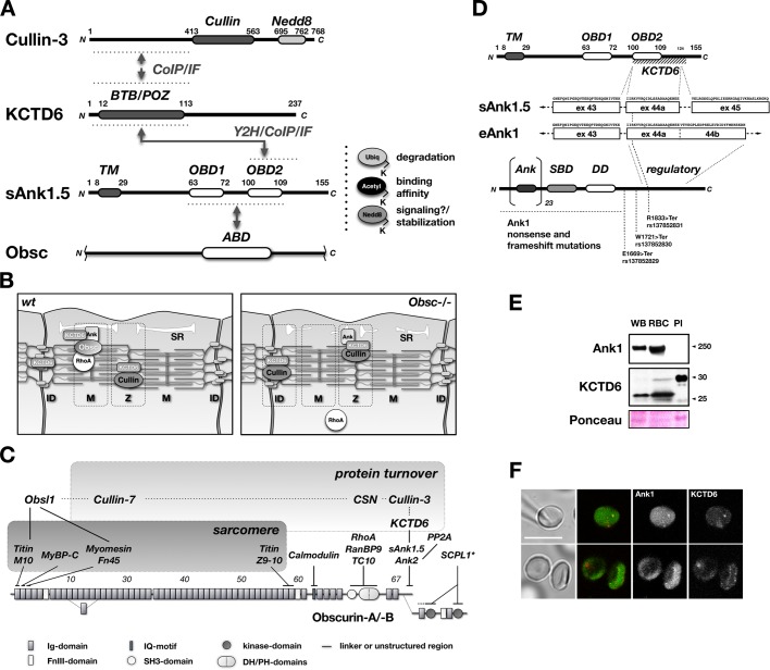
Protein turnover through cullin-3 is tightly regulated by posttranslational modifications, the COP9 signalosome, and BTB/POZ-domain proteins that link cullin-3 to specific substrates for ubiquitylation. In this paper, we report how potassium channel tetramerization domain containing 6 (KCTD6) represents a novel substrate adaptor for cullin-3, effectively regulating protein levels of the muscle small ankyrin-1 isoform 5 (sAnk1.5). Binding of sAnk1.5 to KCTD6, and its subsequent turnover is regulated through posttranslational modification by nedd8, ubiquitin, and acetylation of C-terminal lysine residues. The presence of the sAnk1.5 binding partner obscurin, and mutation of lysine residues increased sAnk1.5 protein levels, as did knockdown of KCTD6 in cardiomyocytes. Obscurin knockout muscle displayed reduced sAnk1.5 levels and mislocalization of the sAnk1.5/KCTD6 complex. Scaffolding functions of obscurin may therefore prevent activation of the cullin-mediated protein degradation machinery and ubiquitylation of sAnk1.5 through sequestration of sAnk1.5/KCTD6 at the sarcomeric M-band, away from the Z-disk-associated cullin-3. The interaction of KCTD6 with ankyrin-1 may have implications beyond muscle for hereditary spherocytosis, as KCTD6 is also present in erythrocytes, and erythrocyte ankyrin isoforms contain its mapped minimal binding site.


Figures

FIGURE 1:
FIGURE 1:
sAnk1.5 interacts with KCTD6 in a tissue-specific manner. (A) Forced yeast two-hybrid identified a region including OBD-2 as minimal binding site for KCTD6. Yeast transformed with either empty pACT2 vector or pACT2-KCTD6 (residues 1–147) together with pLexA-sAnk1.5 constructs expressing small ankyrin regions 29–155, 29–137, 29–99, or 29–63 were grown on SD-LW minimal medium (left). Xgal filter assay (right) indicated a region within sAnk1.5 residues 100–137 as a minimal binding site for sAnk1.5 interaction with KCTD6. (B) Schematic representation of sAnk1.5 domain structure and mapped KCTD6 minimal-binding site. TM, transmembrane domain; OBD, obscurin binding domain. All residues are for human sAnk1.5 (NCBI accession number: NP_065211). (C) Coimmunoprecipitation of either full-length GFP-tagged KCTD6, KCTD6 N-terminus (residues 1–147), or C-terminus (residues 105–237) with HA-tagged sAnk1.5 using cell lysates from transfected Cos-1 or differentiated C2C12 cells (day 7). Full-length KCTD6 displays binding to sAnk1.5 only in lysates from differentiated C2C12 cells, whereas proteins isolated from transfected Cos-1 cells demonstrated only interaction of KCTD6 N-terminus with sAnk1.5. I, input; UB, unbound; B, bound fractions. (D) Schematic representation of KCTD6 domain structure and minimal sAnk1.5 binding site. The minimal binding site to sAnk1.5 encompasses the N-terminal BTB/POZ domain in KCTD6. Results for GST-pulldown assays (Figure S1A), coimmunoprecipitations from transfected Cos-1 or C2C12 cell lysates (C) demonstrate the muscle-specific interaction of sAnk1.5 with full-length KCTD6. All residues are for human KCTD6 (NCBI accession number: NP_699162).
FIGURE 2:
FIGURE 2:
Posttranslational modification of sAnk1.5 affects its interaction with KCTD6. (A) Mutational analysis of putative serine phosphorylation sites and conserved lysine in minimal KCTD6 binding site of sAnk1.5 (residues 100–137). Coimmunoprecipitation of transfected Cos-1 cells expressing GFP-tagged KCTD6 N-terminus with either HA-tagged sAnk1.5 wild-type, Ser-113A, Ser-129A, Ser-113/114A, Ser-113/129A, Ser-113/114/129A, or Lys-101E (Borzok et al., 2007) mutants displayed weaker, but not abrogated, binding of KCTD6 to sAnk1.5 Ser-101E mutant. Full-length KCTD6 shows no binding to sAnk1.5 in Cos-1 cells. (B) HDAC inhibition by TSA triggers binding of sAnk1.5 with full-length KCTD6 in nonmuscle cells. Binding of wild-type and mutant sAnk1.5 (Lys-101E, Glu-121K) to either full-length of C-terminally truncated KCTD6 (residues 1–147) from Cos-1 cells was assessed in absence or presence of 0.5 μg/ml TSA. Mutation of either Lys-101E or Glu-121K did not alter interaction of sAnk1.5 to C-terminally truncated KCTD6. Full-length KCTD6 isolated from COS-1 cells only interacted with sAnk1.5 after incubation of cells with TSA. (C to E) Subcellular localization of endogenous sAnk1.5 (C), KCTD6 (D and E) in heart (C and D) or skeletal muscle cells (E) shows that both proteins localize at the M-band in wild-type cross-striated muscles. (C to E) Scale bar: 10 μm. (F) Acetylation of sAnk1.5 C-terminus. Inhibition of HDAC by TSA leads to increased acetylation of sAnk1.5 as demonstrated by coimmunoprecipitation using an acetyl-lysine antibody (K-Ac; right). Truncation of the sAnk1.5 C-terminus decreases (residues 1–137) or abolishes acetylation (residues 1–63; residues 1–63 Lys-38/46R double mutant).
FIGURE 3:
FIGURE 3:
sAnk1.5 is modified by nedd8 and ubiquitin. Identification of lysine residues important for protein turnover. (A) Protein complementation assay using YFP (split–fluorescent protein assay) demonstrated neddylation of sAnk1.5 in Cos-1 cells. Cos-1 cells transfected with N-terminal YFP-tagged (YN; Zou et al., 2006) nedd8 and C-terminal YFP-tagged (YC) sAnk1.5 (bottom, GFP antibody staining red in overlay) display close association/modification of sAnk1.5 by nedd8 as evidenced by complementation of YFP and restoration of endogenous fluorescence (top). Note that the putative neddylation of sAnk1.5 is readily visible without MG132 treatment. (B) Protein complementation assay as in (A) using YN-ubiquitin and sAnk1.5-YC demonstrated visible modification of sAnk1.5 by ubiquitin only in MG132 treated Cos-1 cells (top). Untreated Cos-1 cells show no detectable ubiquitylation of sAnk1.5, despite expression of constructs (bottom, GFP-antibody red in overlay, DAPI blue in overlay). Scale bar: 20 μm. (C) Analysis of endogenous sAnk1.5 protein degradation in differentiated C2C12 cells using ribosome inhibitor CHX, shows an apparent protein half-life of ∼5 h, comparable with transfected sAnk1.5 in Cos-1 cells (see D). Loading control: β-tubulin. (D) CHX treatment of COS-1 cells transfected with wild-type or mutant sAnk1.5 indicates Lys-38 and Lys-73 as important for ubiquitylation/degradation. Cos-1 cells transfected with HA-tagged wild-type, Lys-38R, Lys-46R, Lys-73R, Lys-105R or Lys-38/73R sAnk1.5 were treated with 10 μg/ml CHX for 6 h. Densitometric quantification of wild-type sAnk1.5 levels indicated a protein half-life of ∼6 h (black bars). Comparison of lysine mutants with the wild-type protein after 6 h of CHX treatment displayed a slight increase of sAnk1.5 for Lys-38R and Lys-73R mutants. The Lys-38/73R double mutant showed significantly increased protein amount (white bar), when compared with wild-type sAnk1.5 after 6 h of treatment. (E) Presence of obscurin C-terminus influences sAnk1.5 degradation. Cos-1 cells transfected with wild-type, Lys-38R, Lys-46R, Lys-73R, Lys-105R or Lys-38/73R sAnk1.5 with or without obscurin C-terminus encompassing the ankyrin binding site (Bagnato et al., 2003; Kontrogianni-Konstantopoulos et al., 2003) were treated with CHX for 6 h. Whereas wild-type sAnk1.5 showed no change in its degradation with or without coexpression of obscurin (black bars), Lys-38R, Lys-73R, Lys-105R and Lys-38/73R mutants show significantly increased sAnk1.5 levels after 6 h of CHX treatment, when coexpressed with obscurin. Similarity of Lys-38R mutant sAnk1.5 levels in presence of obscurin, with amounts of sAnk1.5 Lys-38/73R double mutant (in D) indicate that either Lys-73R mutation, or binding of Lys-73 with obscurin are beneficial for sAnk1.5 protein half-life. (D and E) p values, n values (bottom of each graph), and SEM are displayed. (F) Representation of sAnk1.5 domain layout and posttranslational modification by ubiquitin, nedd8, and acetylation. Lysines in sAnk1.5 and regions responsible for posttranslational modification are indicated (dark shaded = high likelihood of modification). Lysines mutated for protein turnover investigations displayed in bigger letters. Arrows indicate residues influencing turnover.
FIGURE 4:
FIGURE 4:
Association of sAnk1.5 and KCTD6 with E3 ligase cullin-3. (A) KCTD6 associates with the unstructured cullin-3 N-terminus. Coimmunoprecipitation of HA-tagged KCTD6 with GFP-tagged full-length cullin-3, cullin-3 N-terminus (residues 1–384), or cullin-3 C-terminus (residues 384–768) shows binding of KCTD6 to the N-terminal region of cullin-3. (B) Schematic representation of cullin-3 domain structure and minimal KCTD6 binding site as determined in (A). Cullin ligase domain and Nedd8 modification site in cullin-3 are indicated. Residue numbers according to human cullin-3 (NCBI accession number: NP_003581). (C) Coimmunoprecipitation of full-length or sAnk1.5 N-terminus (residues 1–63) demonstrates association of cullin-3 with full-length unmodified sAnk1.5, but not of the sAnk1.5 N-terminus. (A and C) I, input; UB, unbound; B, bound fractions. (D) Schematic representation of ankyrin domain structure and protein region required for association of sAnk1.5 with cullin-3, as demonstrated in (C). (E and F) Immunofluorescence of cardiac (E) and skeletal muscle (F, Dia) display localization of cullin-3 to the sarcomeric Z-disk (arrowheads) as validated by colocalization with sarcomeric α-actinin. Scale bar: 6 μm.
FIGURE 5:
FIGURE 5:
Analysis of KCTD6 developmental and tissue-specific expression pattern. (A) Analysis of endogenous KCTD6 and cullin-3 protein levels in various mouse tissues. High levels of KCTD6 were found in heart (H), kidney (Ki), liver (Li), and stomach (St; top, KCTD6 blot). Ubiquitous low expression was also found in all other analyzed tissues, namely skeletal muscle (Sk), lung (Lu), brain (Br), uterus (Ut), ovaries (Ov), and testes (Te). Note that the KCTD6 antibody cross-reacts with bands at approximately 30 kDa and 25 kDa, with 30 kDa representing full-length KCTD6. Cullin-3 was found to be ubiquitously expressed throughout all analyzed tissue samples (middle). GAPDH was used as loading control (bottom). Asterisk indicates that the liver sample was diluted 1/10 for the KCTD6 blot. (B) Immunoblots of KCTD6 and sAnk1.5 expression in cardiac muscle at P0, P20, and adult stages indicates up-regulation of both proteins during postnatal development. (C) Analysis of sAnk1.5, KCTD6, cullin-3, sarcomeric myosin heavy chain (MyHC), and GAPDH protein levels during C2C12 differentiation. sAnk1.5 and KCTD6 display increased protein levels during C2C12 myotube development, as judged by expression of sarcomeric marker protein MyHC, while cullin-3 remains largely unchanged from undifferentiated myoblast stage (day 0). Note that the larger band in cullin-3 may represent nedd8-modified cullin-3. (D) Analysis of sAnk1.5, KCTD6, cullin-3, and obscurin protein localization in undifferentiated C2C12 myoblasts (day 0) and differentiated C2C12 myotubes. Note that despite overall down-regulation of cullin-3 levels in C2C12 cells (see C), cullin-3 expression levels appear higher in differentiated C2C12 cells (arrowhead in cullin-3 Day 0), when compared with neighboring undifferentiated cells (arrow in cullin-3 bottom). DAPI blue in overlay; sarcomeric marker α-actinin red in overlay. Scale bar: 20 μm.
FIGURE 6:
FIGURE 6:
siRNA knockdown of KCTD6 in NRC. (A) siRNA-mediated knockdown of KCTD6 in NRC leads to reduced expression levels of endogenous KCTD6 when compared with either untransfected (KCTD6 RNAi; right panel, false-color overlay with signal intensity as displayed), or H1-GFP transfected control cells (bottom right panel). (B) Knockdown of endogenous KCTD6 results in increased expression level of endogenous sAnk1.5 (top panel, KCTD6 RNAi), when compared with untransfected (top right panels, false-color overlay with signal intensity as displayed) or H1-GFP–transfected control cells (bottom right panels). (A and B) Scale bar: 20 μm.
FIGURE 7:
FIGURE 7:
Analysis of sAnk1.5, KCTD6, cullin-3, and RhoA expression level and localization in obscurin knockout muscle. (A) Knockout of obscurin leads to changes in sAnk1.5, RhoA, KCTD6, and cullin-3 protein localization. Frozen sections of wild-type and obscurin knockout cross-striated muscle tissues were stained with antibodies directed against sAnk1.5, KCTD6, RhoA, and cullin-3. Prominent M-band localization of sAnk1.5, KCTD6, and RhoA in wild-type muscles appears abrogated in muscles from obscurin knockout animals. sAnk1.5 and RhoA are either diffuse or weakly associated to sarcomeric Z-disks in obscurin knockout muscles, whereas KCTD6 displays prominent Z-disk and intercalated disk association. Intriguingly, prominent localization of cullin-3 to sarcomeric Z-disks remains unchanged. Myomesin, titin-M8, and α-actinin were used as sarcomeric counterstains to show localization of sarcomeric M-band and Z-discs, respectively. Note that the sAnk1.5 staining for obscurin knockout muscles was recorded at higher gain settings compared with the wild-type muscle tissue to emphasize changes to sAnk1.5 localization. Indeed, sAnk1.5 levels were significantly decreased in obscurin knockout muscles (Lange et al., 2009). Scale bar: 10 μm. (B) KCTD6 and cullin-3 localize to the intercalated disk in frozen sections of obscurin cardiac muscle. Cross-sections of muscle from wild-type and obscurin knockout hearts were stained with antibodies raised against KCTD6 and cullin-3 (red in overlay). Plakoglobin (γ-catenin) staining was used as intercalated disk (ID) marker (blue in overlay); F-actin was used to display sarcomeres (green in overlay). Scale bar: 6 μm.
FIGURE 8:
FIGURE 8:
Summary of known and novel obscurin–sAnk1.5 protein interactions and posttranslational modifications. Changes to proteins in obscurin knockouts and a possible link to hereditary spherocytosis. (A) Summary of novel and known protein interactions. Domain structure of obscurin C-terminus, sAnk1.5, KCTD6, and cullin-3 and mapped minimal binding sites are indicated. Amino acid residues are for human sAnk1.5, KCTD6, and cullin-3. OBD, obscurin binding domain; ABD, ankyrin binding domain; TM, transmembrane domain; Obsc, obscurin. Posttranslational modification of sAnk1.5 by nedd8, ubiquitin, and acetylation of lysine residues is indicated. (B) Summary of sAnk1.5 (Ank), KCTD6, RhoA, and cullin protein localization changes between wild-type (left panel) and obscurin knockout heart muscles (right panel). ID, intercalated disk; M, M-band; Z, Z-disk; SR, sarcoplasmic reticulum. (C) Summary of obscurin domain layout and its protein interaction network, with novel and known protein interactions. Proteins involved in sarcomeric structure and protein turnover are grouped. (D) Mapping of KCTD6 site in sAnk1.5 reveals that the exon 44 encoding for the bulk of the binding region is also found in erythrocyte splice variants of Ank1 (eAnk1). Novel and known disease-associated Ank1 mutations that lead to C-terminal truncation of the protein may abrogate putative binding of KCTD6 to ankyrin-1 in erythrocytes. This finding raises the possibility of an involvement of KCTD6 in the development of Ank1-linked hereditary spherocytosis. Domain structure not to scale. Ank, ankyrin repeats; SBD, spectrin binding domain; DD, death domain. (E) Expression of Ank1 and KCTD6 in whole blood (WB), purified red blood cells (RBC), or blood plasma (Pl). The giant ankyrin-1 splice isoforms were detected in whole blood or purified red blood cells. A KCTD6 immunoreactive band at 30 kDa and approximately 25 kDa was detected in purified red blood cells. (F) KCTD6 partially colocalizes in red blood cells with ankyrin-1. Scale bar: 10 μm.

Similar articles

Cited by

References

    1. Alter P, Maisch B. Non-compaction cardiomyopathy in an adult with hereditary spherocytosis. Eur J Heart Fail. 2007;9:98–99. - PubMed
    1. Arendt CS, Hochstrasser M. Eukaryotic 20S proteasome catalytic subunit propeptides prevent active site inactivation by N-terminal acetylation and promote particle assembly. EMBO J. 1999;18:3575–3585. - PMC - PubMed
    1. Bagnato P, Barone V, Giacomello E, Rossi D, Sorrentino V. Binding of an ankyrin-1 isoform to obscurin suggests a molecular link between the sarcoplasmic reticulum and myofibrils in striated muscles. J Cell Biol. 2003;160:245–253. - PMC - PubMed
    1. Bang ML, et al. The complete gene sequence of titin, expression of an unusual approximately 700-kDa titin isoform, and its interaction with obscurin identify a novel Z-line to I-band linking system. Circ Res. 2001;89:1065–1072. - PubMed
    1. Bayon Y, et al. KCTD5, a putative substrate adaptor for cullin3 ubiquitin ligases. FEBS J. 2008;275:3900–3910. - PubMed

Publication types

MeSH terms