. 2012 Aug;26(8):3537-49.
doi: 10.1096/fj.11-197376. Epub 2012 May 9.

Two protein kinase C isoforms, δ and ε, regulate energy homeostasis in mitochondria by transmitting opposing signals to the pyruvate dehydrogenase complex

Affiliations

Two protein kinase C isoforms, δ and ε, regulate energy homeostasis in mitochondria by transmitting opposing signals to the pyruvate dehydrogenase complex

Jianli Gong et al. FASEB J. 2012 Aug.

Abstract

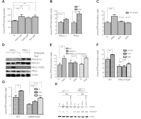
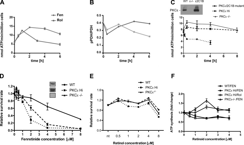
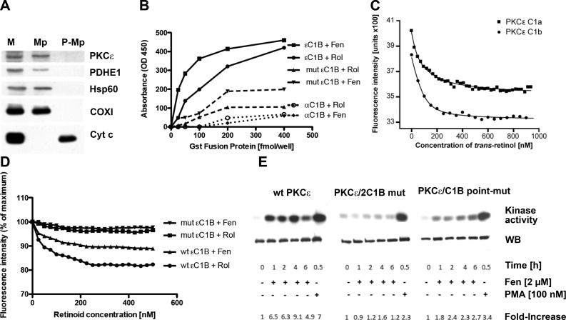
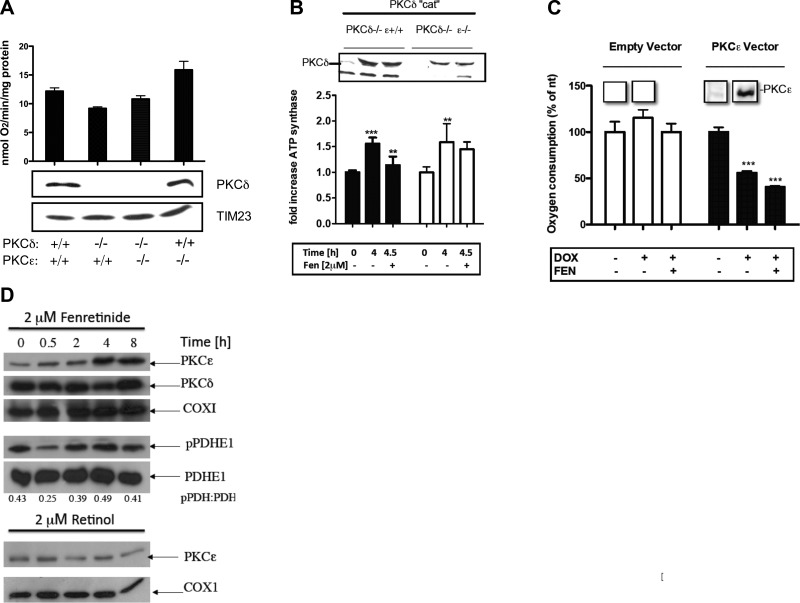
Energy production in mitochondria is a multistep process that requires coordination of several subsystems. While reversible phosphorylation is emerging as the principal tool, it is still unclear how this signal network senses the workloads of processes as different as fuel procurement, catabolism in the Krebs cycle, and stepwise oxidation of reducing equivalents in the electron transfer chain. We previously proposed that mitochondria use oxidized cytochrome c in concert with retinol to activate protein kinase Cδ, thereby linking a prominent kinase network to the redox balance of the ETC. Here, we show that activation of PKCε in mitochondria also requires retinol as a cofactor, implying a redox-mechanism. Whereas activated PKCδ transmits a stimulatory signal to the pyruvate dehdyrogenase complex (PDHC), PKCε opposes this signal and inhibits the PDHC. Our results suggest that the balance between PKCδ and ε is of paramount importance not only for flux of fuel entering the Krebs cycle but for overall energy homeostasis. We observed that the synthetic retinoid fenretinide substituted for the retinol cofactor function but, on chronic use, distorted this signal balance, leading to predominance of PKCε over PKCδ. The suppression of the PDHC might explain the proapoptotic effect of fenretinide on tumor cells, as well as the diminished adiposity observed in experimental animals and humans. Furthermore, a disturbed balance between PKCδ and PKCε might underlie the injury inflicted on the ischemic myocardium during reperfusion. dehydrogenase complex.


Figures

Figure 1.
Figure 1.
Fenretinide as coactivator of PKCδ. A) Capacity of fenretinide to accelerate the ATP synthase rate of WT MEFs equals that of retinol. WT MEFs were treated for 30 min with either 1 or 2 μM fenretinide (FEN) or 2 μM retinol (Rol), or left untreated. Pyruvate/malate-driven rates of ATP synthesis were measured as described in Materials and Methods and expressed as nanomoles ATP per minute per milligram protein or per million cells. ATP synthesis increased by 28, 25, or 28%, respectively (n=6). One of 3 independent experiments is shown. **P < 0.01, ***P < 0.001. B) Failure to stimulate ATP synthesis in PKCδ−/− MEFs indicates dependence on PKCδ; reintroduction of PKCδ restores responsiveness to fenretinide. PKCδ−/− MEFs, and PKCδ−/− MEFs reconstituted with full-length PKCδ by transfection, were treated with fenretinide and analyzed for ATP production as in A. The response of PKCδ−/− MEFs was statistically insignificant, whereas reintroduction of PKCδ reestablished response to fenretinide (45% increase, n=3). One of 4 independent experiments is shown. **P < 0.01. C) Expression of a mutant PKCδ gene deficient in retinoid-binding sites fails to rescue the PKCδ phenotype. This block is bypassed by phorbol ester (PMA). PKCδ was converted to a retinol-nonbinding form by genetic exchange of both endogenous zinc-finger domains for the PKCα C1B domain. The reengineered full-length gene was expressed in PKCδ−/− MEFs. Responsiveness to retinoids (2 μM), or PMA (100 nM) was determined as described in A. Neither retinol nor fenretinide elicited a cofactor response commensurate with WT PKCδ responses shown in A and B. A 35% increase in ATP synthesis (n=4) was observed with PMA, which bypasses the requirement for retinoid cofactor. ***P < 0.001. D) Fenretinide stimulates autophosphorylation of threonine-505 of PKCδ, indicating conversion to active enzyme. The E1 subunit of PDH is dephosphorylated, indicating PDH activation. WT and PKCδ−/− MEFs were stimulated with 2 μM fenretinide as in A. Mitochondrial proteins were separated by SDS-PAGE and analyzed by immunoblotting for the proteins indicated. Phosphorylation of T 505 of PKCδ and dephosphorylation of PDH E1 indicated activation of both enzymes, and this was prevented by rottlerin (Rott). PDH was not dephosphorylated in PKCδ−/− MEFs. One of 3 independent experiments is shown. Direct immunoblotting of PKCδ stained a contaminating band. This was eliminated by prior enrichment of PKCδ with immunoprecipitation (not shown). E) Fenretinide-mediated up-regulation of oxidative phosphorylation is ablated by p66Shc gene knockout, and it is restored by reintroduction of the intact gene. WT MEFs, MEFs with a defective p66Shc gene, and p66Shc−/−MEFs reconstituted with intact p66Shc gene were treated with fenretinide and analyzed as in A. ATP synthesis increased by 30% in WT MEFs, was not enhanced in the knockout MEFs, but was enhanced by 30% after reintroduction of p66Shc (P < 0.002 for both responders, n=4). One of 3 independent experiments is shown. **P < 0.01. F) Fenretinide-mediated up-regulation of oxidative phosphorylation is abolished in MEFs expressing the Y332F mutant PKCδ, defective in its ability to bind p66Shc. WT MEFs and PKCδ−/− MEFs reconstituted with the mutated PKCδ Y332F gene were treated with 2 μM fenretinide and analyzed as in A. WT cells responded by a 25% increase in ATP synthesis (P < 0.01, n=4), but MEFs carrying the defective PKCδ did not. The block was partially overridden by stimulation with PMA. One of 6 independent experiments is shown. **P < 0.01. G) Fenretinide-mediated up-regulation of oxidative phosphorylation is abolished in MEFs expressing the E132Q/E133Q mutant of p66Shc incapable of binding cytochrome c. WT MEFs and p66Shc−/− MEFs reconstituted with the mutated p66ShcQQ gene were treated with 2 μM fenretinide and analyzed as in A. WT cells responded by a 25% increase in ATP synthesis (P < 0.01), but responses of MEFs carrying the mutated QQ gene were attenuated. PMA partially overrode this block (35% increase, P < 0.14). One of 6 independent experiments is shown. **P < 0.01. H) PKCδ signaling is defective in mitoplasts deprived of cytochome c, and is restored by introduction of oxidized cytochrome c protein in combination with fenretinide (left panel); mitoplasts of PKCδ−/− MEFs fail to respond to cytochrome c and fenretinide (right panel). Mitochondria (mt) and mitoplasts (Mp) of cytochrome c-knockdown (cyt c−/−) MEFs or cytochrome c-knockdown/PKCδ-deficient (cyt c−/− PKCδ−/−) cells were prepared as described in Materials and Methods. Mitoplasts were reconstituted with 25 mM oxidized cytochrome c (cyt cox) with and without fenretinide for 10 min at 37°C, or left untreated (nt). Mitochondrial proteins were separated by SDS-PAGE and analyzed by immunoblot for PDH E1 content and phosphorylation status. Time 23 was used as a loading control. Dephosphorylation was observed solely in mitoplasts treated with the combination of cytochrome c and fenretinide (6-fold reduction by densitometry). All other preparations, including the mitochondria and mitoplasts devoid of PKCδ, did not yield dephosphorylated PDH E1 species. One of two repeat experiments is shown.
Figure 2.
Figure 2.
The inhibitory effects of late-acting fenretinide are mediated by PKCε. A, B) Fenretinide elicits biphasic responses, as shown for ATP synthesis (A) and PDH phosphorylation status (B). WT MEFs were stimulated with 2 μM retinol (Rol) or fenretinide (Fen) for indicated time periods and analyzed for ATP synthesis as described in Fig. 1A. In parallel, mitochondrial proteins were separated by SDS gel electrophoresis and analyzed for PDH E1 content and phosphorylation status by immunoblotting. Ratios of PDH: phosphoPDH E1 were determined densitometrically (B). The monophasic retinol response contrasts with the biphasic response to fenretinide (A). The late-phase decline is characteristic of fenretinide and is less pronounced with retinol. One of 3 independent experiments is shown. C) PKCε phenotype influences the changes in ATP synthesis rates elicited by fenretinide. MEFs expressing elevated levels of PKCε (PKCεHi), MEFs expressing a defective PKCε gene (PKCε−/−) and PKCε−/− cells reconstituted with a mutated PKCε gene lacking intact retinoid binding sites (PKCε/2C1B domain-exchange mutant) were treated and analyzed as in A. Expression levels were monitored by immunoblot (inset). PKCε gene ablation abolished the late-phase inhibition but permitted the short-term up-regulation of ATP synthesis. Conversely, PKCε overexpression (6-fold over WT levels) abolished the short-term ATP up-regulation and accelerated fenretinide-mediated ATP suppression. Expression of mutant PKCε, defective in the ability to bind retinoids, in PKCε−/− MEFs did not restore fenretinide mediated suppression. One of 3 independent experiments is shown. D, E) Fenretinide-mediated cell death is influenced by the PKCε phenotype. WT, PKCε Hi, and PKCε−/− MEFs were cultured in serum-free medium and treated with indicated concentrations of fenretinide (D) or retinol (E) for 24 h. Insets show expression levels by immunoblotting. Relative survival of cells was determined as described in ref. . Ablation of PKCε conferred resistance to fenretinide, whereas overexpression of PKCε conferred increased sensitivity. Up to 4 μM retinol preserved the viability of all three cell types. Above 4 μM, cell viability declined due to toxicity. One of 6 independent experiments is shown. ***P < 0.01. F) Optimal dose-responses of fenretinide and retinol are in the range of 1 to 2 μM. WT, PKCε−/−, and PKCε overexpressing (PKCε Hi) MEFs were stimulated for 30 min with the doses of retinoids shown, and rates of ATP synthesis were determined. Fold changes ± se relative to cells treated with vehicle are shown. In all 4 dose-response curves, differences between treatments with vehicle and 2-μM retinoids reached statistical significance (P < 0.014 or better).
Figure 3.
Figure 3.
PKCε residing in the mitochondrial matrix binds retinol and fenretinide; cofactor binding to the activation domain is required for in situ conversion to active enzyme. A) PKCε resides in the matrix. Mouse liver mitochondria were treated with digitonin to permeabilize the outer membrane to generate mitoplasts (Mp; ref. 28). Proteins of mitochondria (M), mitoplasts, and postmitoplast supernates (P-Mp) were separated by SDS-PAGE and analyzed by immunoblotting. Western blots reveal the presence of PKCε in intact mitochondria and mitoplasts along with matrix proteins: PDH E1, Hsp60, and COXI. Mitoplasts released proteins associated with the intermembrane space, including cytochrome c (Cyt c). PKCε was notably absent from postmitoplast supernates. B) ELISA assays using solid-phase retinol (Rol) or fenretinide (Fen) indicate specific interaction of GST-PKCεC1B fusion protein with both retinoids. Ablation of 2 contact sites in the retinol-binding pocket attenuates binding. Control GST-PKCαC1B fusion protein did not bind retinoids as expected (32). One of 3 independent experiments is shown. C, D) PKCε contains 2 retinoid-binding sites associated with zinc-finger domains. Retinoid-binding assays were based on quenching of tryptophane fluorescence (23, 71). Both εC1A and εC1B recombinant zinc-finger proteins bound retinol with high affinity (Kd=87 and 24 nM; C). Although fenretinide quenched tryptophane fluorescence emission with lower amplitude, its nominal binding affinity for εC1B (Kd=60 nM) is in range with that of retinol. Fluorescence quenching is abolished in the double-mutant GST-PKCεC1B fusion protein, affirming specificity of retinoid binding (D). E) Activation of PKCε in mitochondria depends on binding of retinoid cofactor. MEFs expressing the WT (left panel), the domain-exchange mutated (middle panel), or the double point-mutated PKCε gene (right panel) were rendered quiescent in serum-free and retinoid-free medium. They were reactivated by FBS in the presence of fenretinide. PKCε was immunoprecipitated from mitochondrial lysates and assayed for phosphotransferase activity for the histone substrate. Note that in the presence of FBS 2 μM fenretinide does not kill MEFs (data not shown). Blots show the autoradiographs of histone phosphorylation, and immunoblots of PKCε levels below. The fold increases are presented as the ratios of phospho-histone of activated vs. quiescent cells, normalized for the PKCε amounts recovered. WT PKCε was on average 4 times as active compared to two retinoid binding-deficient PKCε constructs.
Figure 4.
Figure 4.
Respiration is controlled by the balance between PKCδ and PKCε. A) Baseline levels of respiration are influenced by PKCδ and PKCε phenotypes. WT, PKCδ−/−, PKCε−/−, and PKCδ−/− PKCε−/− MEFs were rendered quiescent by culturing in serum-free medium and analyzed for baseline oxygen consumption without reactivation. MEFs lacking a functional PKCδ gene displayed depressed baseline level of respiration compared to WT MEFs. Conversely, ablation of PKCε resulted in elevated respiration. The dual δ,ε-knockout MEFs equaled WT cells in oxygen consumption. Tim23 levels were comparable in all 4 cell types, suggesting equal amounts of mitochondria. This was confirmed by mitotracker staining (data not shown). PKCε-knockout MEFs had no more PKCδ in their mitochondria than WT cells, implicating the missing PKCε gene in negative regulation of respiration. B) Constitutively active PKCδ stimulates respiration, which is attenuated by fenretinide-assisted PKCε activation. WT PKCδ cat was placed under a tetracycline-inducible promoter and transfected into either PKCδ−/− PKCε+/+ or PKCδ−/− PKCε−/− MEFs by a lentiviral vector (26, 27). Expression was induced by doxycyclin. Presence of PKCδ cat in mitochondria was monitored by immunoblot (inset). Increased ATP synthesis (1.5-fold) coincided with PKCδ-cat appearance after 4 h of induction in both cell lines. The addition of 2 μM fenretinide for the last 30 min decreased ATP synthesis to near baseline level in the former but not PKCε-knockout MEFs, indicating that suppression of ATP synthesis was dependent on PKCε. **P < 0.01, ***P < 0.001. C) Activation of PKCε leads to diminished respiration. The full-length PKCε gene under inducible promoter was expressed in PKCε−/− cells by lentiviral transfection. Induction of PKCε expression by doxocycline was observed after 4 h (see inset showing PKCε immunoblot of mitochondrial proteins) and caused 50% reduction of oxygen consumption. The addition of fenretinide elicited further 15% reduction. MEFs transfected with empty vector did not display suppression of ATP synthesis. ***P < 0.001. D) Fenretinide causes selective accumulation of PKCε in mitochondria. WT MEFs were stimulated with 2 μM fenretinide for the indicated periods of time. Mitochondria were isolated, and their proteins were separated by SDS-PAGE and analyzed by immunoblotting for the indicated proteins. ATP synthesis rates and phosphorylation status of PDHE1 were determined as described in Fig. 1A. Scans of similar immunoblots displayed here are presented in Fig. 2A, B. Fenretinide stimulated a progressive increase in the amount of PKCε present in mitochondria. PKCδ remained at a constant level (top panels). The late-phase PKCε accumulation correlated with PDH E1 hyperphosphorylation, indicating loss of PDHC activity. The ratios of phospho-PDH to PDH E1 were determined by densitometry. COXI was used as a loading control (middle panels). The treatment of cells with 2 μM retinol did not result in accumulation of PKCε in mitochondria over the 8-h period (bottom panels). Experiment shown is representative of 3 repeats.

References

    1. Holness M. J., Sugden M. C. (2003) Regulation of pyruvate dehydrogenase complex activity by reversible phosphorylation. Biochem. Soc. Trans. 31, 1143–1151 - PubMed
    1. Bowker-Kinley M. M., Davis W. I., Wu P., Harris R. A., Popov K. M. (1998) Evidence for existence of tissue-specific regulation of the mammalian pyruvate dehydrogenase complex. Biochem. J. 329, 191–196 - PMC - PubMed
    1. Huang B., Gudi R., Wu P., Harris R. A., Hamilton J., Popov K. M. (1998) Isoenzymes of pyruvate dehydrogenase phosphatase. DNA-derived amino acid sequences, expression, and regulation. J. Biol. Chem. 273, 17680–17688 - PubMed
    1. D'Aurelio M., Pallotti F., Barrientos A., Gajewski C. D., Kwong J. Q., Bruno C., Beal M. F., Manfredi G. (2001) In vivo regulation of oxidative phosphorylation in cells harboring a stop-codon mutation in mitochondrial DNA-encoded cytochrome c oxidase subunit I. J. Biol. Chem. 276, 46925–46932 - PubMed
    1. Acin-Perez R., Salazar E., Kamenetsky M., Buck J., Levin L. R., Manfredi G. (2009) Cyclic AMP produced inside mitochondria regulates oxidative phosphorylation. Cell Metab. 9, 265–276 - PMC - PubMed

Publication types

MeSH terms

LinkOut - more resources