. 2012:(183):37-83.
doi: 10.3897/zookeys.183.2556. Epub 2012 Apr 19.

A review of the North American genus Epimartyria (Lepidoptera, Micropterigidae) with a discussion of the larval plastron

Affiliations

A review of the North American genus Epimartyria (Lepidoptera, Micropterigidae) with a discussion of the larval plastron

Donald R Davis et al. Zookeys. 2012.

Abstract

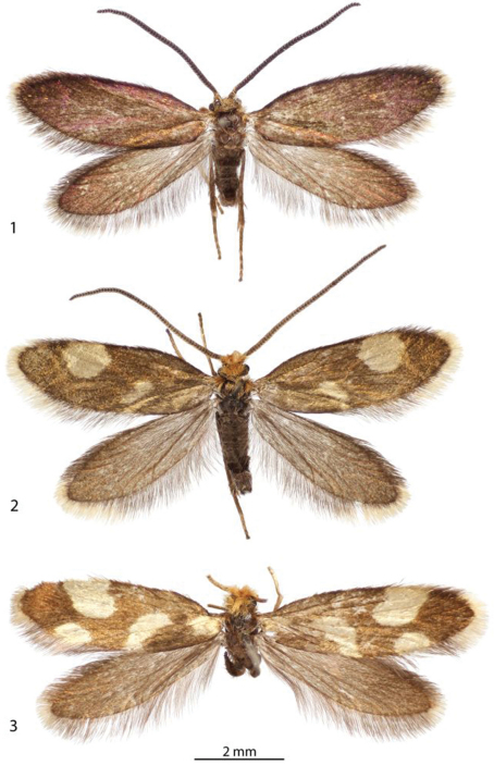
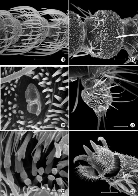
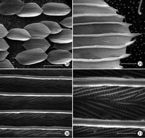
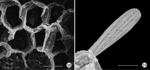
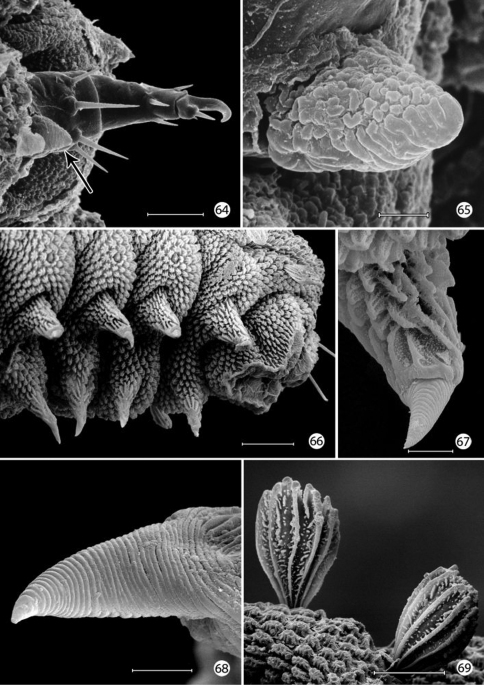
The indigenous North American micropterigid genus Epimartyria Walsingham,1898 is revised. Three species are recognized, including Epimartyria auricrinella Walsingham, 1898 which occurs widely over much of the northeastern United States and Canada, a new species, Epimartyria bimaculella Davis & Landry from northwestern United States and Canada, and Epimartyria pardella (Walsingham, 1880) from northern California to northern Oregon. The larva of Epimartyria auricrinella is described in detail, supplemented with illustrations of the external structure of the larval integument. The larval plastron is described and illustrated for Epimartyria, and this is compared with the plastrons of Neomicropteryx Issiki, 1931 and Micropterix Hübner, 1825. COI barcode sequences show that the three species are genetically distinct, congruent with morphological differences. Marked haplotype divergence within some Epimartyria auricrinella populations appears to be unrelated to morphology, geography or phenology.

Keywords: DNA barcodes; Distribution; genital morphology; larval morphology; plastron.


Figures

Figures 1–3.
Figures 1–3.
Adults. 1 ♂, Epimartyria auricrinella,  (4.9 mm) Canada: Quebec 2 ♂, Epimartyria bimaculella  (5.5 mm) Holotype, Canada: British Columbia 3 ♀, Epimartyria pardella (5.5 mm) USA: California. (Forewing length in parentheses).
Figures 4–9.
Figures 4–9.
Adults and habitat. 4–5 Epimartyria auricrinella, at Lac Brûlé, Québec, 30 Jun 1997, ca 0700 hrs. on dewy Solidago leaf 6–7 Epimartyria bimaculella 6 at Washington, Olympic National Park, Hoh Rainforest Road, 22 Jun 2010 (photo by Zeller-Lukashort) 7 at British Columbia, Vancouver area, Belcarra, 24 May 2009, ca 1000 hrs (photo by Holden) 8 Epimartyria pardella, California, Redwood National Park, Gold Bluffs State Beach, Fern Canyon 9 Habitat, clump of the liverwort Bazzania trilobata at Lac Brûlé, Québec in which larvae of Epimartyria auricrinella were found.
Figures 10–11.
Figures 10–11.
Habitats of Epimartyria 10 Swampy forest at Lac Brûlé, Quebec where larvae and numerous adults of Epimartyria auricrinella were collected 11 Douglas fir forest where adults of Epimartyria bimaculella were observed swarming around the ferns (photo by Zeller-Lukashort).
Figure 12a.
Figure 12a.
Neighbour-joining tree of genetic distances (K2P model) for cytochrome c oxidase I (COI) in species of Epimartyria (total = 44 specimens). End-branch labels are specimen Sample IDs followed by the geographic area in parentheses: BC = British Columbia; CA = California; MI = Michigan; QC = Quebec; TN = Tennessee; WA = Washington. Sequence lengths are 658bp unless otherwise indicated (xn in square brackets indicates the number of ambiguous positions). Distinct haplotypes are designated by a capital letter and digit.
Figure 12b.
Figure 12b.
Strict consensus tree of three most parsimonious trees (length = 65, CI = 0.877, RI = 0.857) based on 11 unique DNA barcode haplotypes in species of Epimartyria. End-branch alphanumeric labels are specimen SampleIDs with haplotype designations (A1, A2, etc.). Numbers above branches are bootstrap values (1000 replicates) / Bremer support values.
Figure 12c.
Figure 12c.
Haplotype network for 10 distinct haplotypes detected in two species of Epimartyria (7 for Epimartyria auricrinella, 4 for Epimartyria bimaculella). Circles are labelled with the haplotype name (capital letter), and the number of specimens per haplotype; lower case letters refer to localities indicated on the distribution map (Fig. 32). The single sequence of Epimartyria pardella, which separated out, is not shown.
Figures 13–17.
Figures 13–17.
Epimartyria pardella, Adult morphology 13 Head (cl: clypeus; ga: galea; la: labrum; md: mandible) (0.5 mm) 14 Head, ventral view ( pp: proximal prelabium; dp: distal prelabium; lp: labial palpus) 15 right mandible 16 Wing venation, USNM slide 16613. 17 Legs (1.0 mm). (Scale lengths in parentheses).
Figures 18–23.
Figures 18–23.
Epimartyria auricrinella, Adultmorphology 18 Flagellomeres with ascoid sensilla (20 µm) 19 Flagellomere with multiporus sensillum placodeum (20 µm) 20 Detail of multiporus sensillum placodeum in Fig. 19 (2 µm) 21 Apical segment of labial palpus with distal organ vom Rath (20 µm). 22 Sensilla of organ vom Rath (2 µm) 23 Mesothorcic pretarsus (20 µm). (Scale lengths in parentheses).
Figures 24–27.
Figures 24–27.
Epimartyria auricrinella, Adultmorphology 24 Mesothorcic pretarsus (20 µm) 25 Detail of pseudempodium of pretarsus (2 µm) 26 Arolium (5 µm) 27 Detail of surface of arolium (1 µm). (Scale lengths in parentheses).
Figures 28–31.
Figures 28–31.
Epimartyria auricrinella, Forewing scale structure 28 Dorsal forewing scales from discal cell (40 µm) 29 Apical margin of scale in Fig. 28 (2 µm) 30 detail of Fig. 29 (2 µm) 31 Detail of Fig. 30 (1 µm). (Scale lengths in parentheses).
Figure 32.
Figure 32.
Distribution of Epimartyria species. Alphanumeric designations refer to haplotypes shown in the haplotype network of Fig. 12c.
Figures 33–39.
Figures 33–39.
Epimartyria auricrinella, larval morphology 33 Chaetotaxy; shaded area indicates extent of epidermal plastron34 Head, dorsal view(M: medial seta) 35 Head, ventral view (AT: anterior arm of tentorium) 36 Ventralview of maxilla and labrum 37 Head, lateral view 38 Labrum, dorsal view 39 Mandible.
Figures 40–45.
Figures 40–45.
Epimartyria auricrinella, larval morphology 40 Head, dorsal view (100 µm) 41 head, anterior view (100 µm) 42 Scutate cuticular outgrowths from head-prothoracic fold (of Fig. 41) (10 µm) 43 Apex of antenna (10 µm). 44 Stemmata, 5 total (25 µm) 45 Ventral view of maxilla and labium (20 µm). (Scale lengths in parentheses).
Figures 46–51.
Figures 46–51.
Epimartyria auricrinella, larval morphology 46 Apical sensilla of maxillary palpus (5 µm) 47 Labial palpi and opening of labial salivary gland (100 µm) 48 Thoracic legs (arrow: tactile vesicle of coxa) (200 µm) 49 Mesothoracic leg (arrow: tactile vesicle of coxa) (25 µm) 50 Abdominal prolegs, segments 1–2 (100 µm) 51 Anal prolegs (100 µm). (Scale lengths in parentheses).
Figures 52–57.
Figures 52–57.
Epimartyria auricrinella, larval morphology 52 Abdominal segments 1–4, lateral view, showing sculptured epicuticle of dorsal half and plastron region (shaded area) of lower half (arrow indicates spiracle) (200 µm) 53 Spiracle, apical view (5 µm) 54 Spiracle, lateral view (5 µm) 55 Plastron of lateral surface of abdomen with numerous, irregular micropapillae (10 µm) 56 Detail of fig. 55 showing parallel rows of microtubercules extending between micropapillae (1 µm) 57 View looking down on microtubules in fig. 56 showing cuticular openings between rows (1 µm). (Scale lengths in parentheses).
Figures 58–59.
Figures 58–59.
Epimartyria auricrinella, larval morphology 58 Honeycombed chambers of abdominal exocuticle with pellicle removed, in dorsal half (dorsal to spiracle) of abdominal segment 4 (10 µm) 59 Abdominal seta D1 showing longitudinal ridges (20 µm). (Scale lengths in parentheses).
Figures 60–63.
Figures 60–63.
Neomicropteryx nipponensis, larval morphology 60 Plastron from lateral surface of abdomen with numerous, irregular micropapillae (10 µm) 61 Detail of Fig. 55 showing parallel rows of microtubercules extending between micropapillae (0.5 µm) 62 View looking down on microtubules in fig. 61 showing cuticular openings between rows (0.5 µm) 63 Head, anterior view (100 µm). (Scale lengths in parentheses).
Figures 64–69.
Figures 64–69.
Micropterix species (England), larval morphology 64 Prothoracic leg (arrow: tactile vesicle of coxa) (50 µm) 65 Detail of tactile vesicle in fig. 64 (10 µm) 66 Abdominal segments 5–10 showing prolegs 5–8 and sucker-like anal proleg (100 µm) 67 Abdominal proleg (20 µm) 68 Detail of apex of abdominal proleg (10 µm) 69 Dorsal abdominal setae (50 µm). (Scale lengths in parentheses).
Figures 70–73.
Figures 70–73.
Micropterix species (England), larval morphology 70 Lateral plastron surface of abdomen showing micropapillae around broken scale base (20 µm) 71 Detail of plastron Fig. 70 showing cuticular openings between micropapillae (10 µm) 72 Detail of fig. 71 (5 µm) 73 Detail ofcuticular openings in Fig. 71 (2 µm). (Scale lengths in parentheses).
Figures 74–80.
Figures 74–80.
Epimartyria auricrinella, Genitalic morphology 74–78 Male, USNM slides 16615, 34372 74 Genital capsule, ventral view (0.5 mm); J: juxta (medial plate) 75 Sternum X (gnathos) 76 Aedeagus (G: gonopore (phallotreme) 77 Genital capsule, lateral view (Un: uncus, (tergum X) 78a Valva, lateral view, inner side (BP: basal process), slide USNM, 34372, Ottawa, Ontario 78b slide MIC5762, Lac Brûlé, Quebec 78c slide MIC5764, Wilderness State Park, Michigan 78d slide MIC5761, Lac Brûlé, Quebec 79–80 Female, USNM slide 17501, Mt. Albert, Quebec 79 Oviscape, lateral view (Ut: utriculus) (0.5 mm) 80 Genital sclerite, ventral view. (Scale lengths in parentheses).
Figures 81–87.
Figures 81–87.
Epimartyria bimaculella, Genitalic morphology 81–85 Male, USNM slide 18410, Fraser Mill, British Columbia 81 Genital capsule, ventral view (0.5 mm) 82 Sternum X (gnathos) 83 Aedeagus 84 Genital capsule, lateral view 85 Valva 86–87 Female, USNM slide 33919, Fraser Mill, British Columbia 86 Oviscape, lateral view (0.5 mm) 87 Genital sclerite, ventral view. (Scale lengths in parentheses).
Figures 88–96.
Figures 88–96.
Epimartyria pardella, Genitalic morphology 88–92 Male, USNM slide 16613, Arcata, California 88 Genital capsule, ventral view (0.5 mm) 89 Sternum X (gnathos) 90 Aedeagus 91 Genital capsule, lateral view 92 Valva 93–96 Female, DRD slide 4528, Kneeland, California 93 Oviscape, lateral view (0.5 mm) 94 Genital sclerite, ventral view 95 Genital sclerite, lateral view 96 Ductus spermathecae, showing variation of vesicle position. (Scale lengths in parentheses).

References

    1. Carter DJ, Dugdale JS. (1982) Notes on collecting and rearing Micropterix (Lepidoptera: Micropterigidae) larvae in England. Entomologist’s Gazette 33: 43-47.
    1. Davis DR. (1975) A review of the West Indian moths of the family Psychidae with descriptions of new taxa and immature stages. Smithsonian Contributions to Zoology 188: 1-66. doi: 10.5479/si.00810282.188 - DOI
    1. Davis DR. (1983) Micropterigidae. In: RW Hodges et al. (Eds) Check List of the Lepidoptera of America North of Mexico. EW Classey Ltd and the Wedge Entomological Research Foundation, London, 5–7.
    1. Davis DR. (1986) A New Family of Monotrysian Moths from Austral South America (Lepidoptera: Palaephatidae), with a Phylogenetic Review of the Monotrysia. Smithsonian Contributions to Zoology 434: 1–202. [599 Figs, 15 maps, 2 tables]
    1. Davis DR. (1987) Micropterigidae, Eriocraniidae. In: FW Stehr. (Ed). Immature insects. Volume 1, Kendall/Hunt Publishing, Dubuque, Iowa: 341-343.

LinkOut - more resources