Two cases of female hydrocele of the canal of nuck
- PMID: 22574075
- PMCID: PMC3346837
- DOI: 10.3345/kjp.2012.55.4.143
Two cases of female hydrocele of the canal of nuck
Abstract
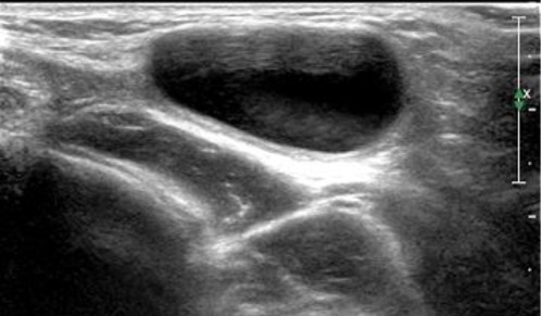
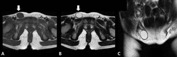
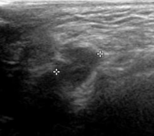
The processus vaginalis within the inguinal canal forms the canal of Nuck, which is a homolog of the processus vaginalis in women. Incomplete obliteration of the processus vaginalis causes indirect inguinal hernia or hydrocele of the canal of Nuck, a very rare condition in women. Here, we report 2 cases of hydrocele of the canal of Nuck that were diagnosed with ultrasonography in both cases and magnetic resonance imaging in 1 case to confirm the sonographic diagnosis. High ligation and hydrocelectomy were conducted in both patients. In 1 patient, 14 months later, the occurrence of contralateral inguinal hernia was suspected, but did not require surgery. The other patient had a history of surgery for left inguinal hernia 11 months before the occurrence of right hydrocele of the canal of Nuck. In both cases, the occurrence of an inguinal hernia on the contralateral side was noted.
Keywords: Canal of Nuck; Hydrocele; Inguinal hernia; Magnetic resonance imaging; Ultrasonography.
Figures
References
-
- Anderson CC, Broadie TA, Mackey JE, Kopecky KK. Hydrocele of the canal of Nuck: ultrasound appearance. Am Surg. 1995;61:959–961. - PubMed
-
- Schwartz A, Peyser MR. Nuck's hydrocele (hydrocele muliebris) Int Surg. 1975;60:91–92. - PubMed
-
- Kim SH, Seo IY, Cho HJ, Ku1 YM, Kim KH, Ahn CH, et al. Hydrocele of the Canal of Nuck. J Korean Surg Soc. 2008;74:396–398.
-
- Park SJ, Lee HK, Hong HS, Kim HC, Kim DH, Park JS, et al. Hydrocele of the canal of Nuck in a girl: ultrasound and MR appearance. Br J Radiol. 2004;77:243–244. - PubMed
-
- Haynes JH. Inguinal and scrotal disorders. Surg Clin North Am. 2006;86:371–381. ix. - PubMed
LinkOut - more resources
Full Text Sources