

Auditory resting-state network connectivity in tinnitus: a functional MRI study
- PMID: 22574141
- PMCID: PMC3344851
- DOI: 10.1371/journal.pone.0036222
Auditory resting-state network connectivity in tinnitus: a functional MRI study
Abstract
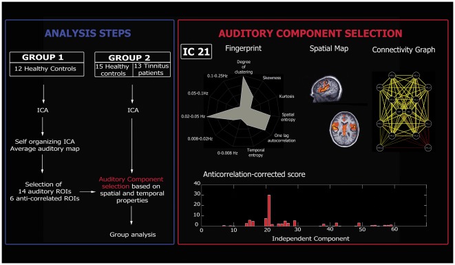
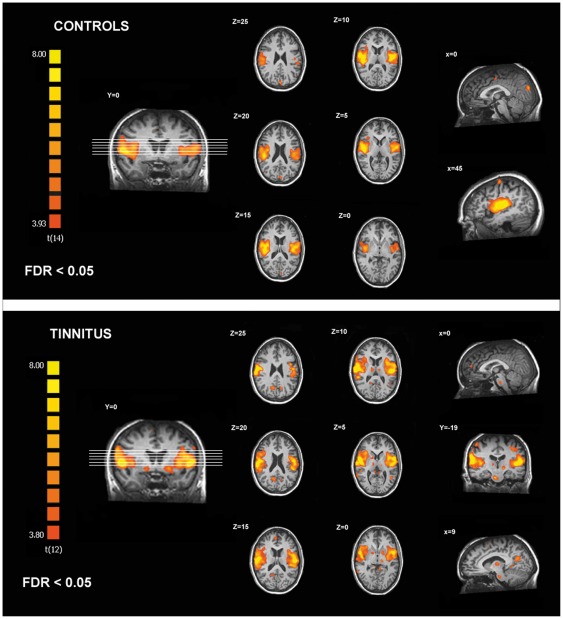
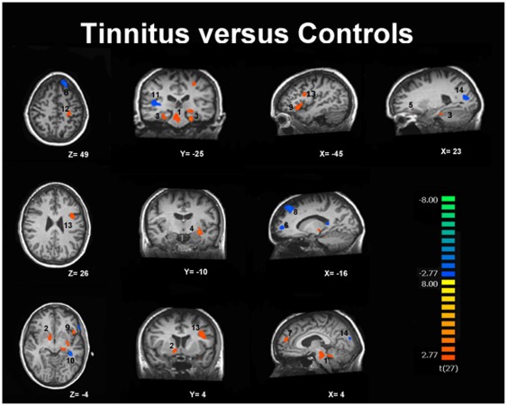
The underlying functional neuroanatomy of tinnitus remains poorly understood. Few studies have focused on functional cerebral connectivity changes in tinnitus patients. The aim of this study was to test if functional MRI "resting-state" connectivity patterns in auditory network differ between tinnitus patients and normal controls. Thirteen chronic tinnitus subjects and fifteen age-matched healthy controls were studied on a 3 tesla MRI. Connectivity was investigated using independent component analysis and an automated component selection approach taking into account the spatial and temporal properties of each component. Connectivity in extra-auditory regions such as brainstem, basal ganglia/NAc, cerebellum, parahippocampal, right prefrontal, parietal, and sensorimotor areas was found to be increased in tinnitus subjects. The right primary auditory cortex, left prefrontal, left fusiform gyrus, and bilateral occipital regions showed a decreased connectivity in tinnitus. These results show that there is a modification of cortical and subcortical functional connectivity in tinnitus encompassing attentional, mnemonic, and emotional networks. Our data corroborate the hypothesized implication of non-auditory regions in tinnitus physiopathology and suggest that various regions of the brain seem involved in the persistent awareness of the phenomenon as well as in the development of the associated distress leading to disabling chronic tinnitus.
Conflict of interest statement
Figures
References
-
- Moller . Textbook of Tinnitus. New York: Springer; 2011.
-
- Eggermont JJ, Roberts LE. The neuroscience of tinnitus. Trends Neurosci. 2004;27:676–682. - PubMed
-
- Adjamian P, Sereda M, Hall DA. The mechanisms of tinnitus: perspectives from human functional neuroimaging. Hear Res. 2009;253:15–31. - PubMed
-
- Barrs D, Brackmann D. Translabyrinthine nerve section: effect on tinnitus. The Journal of Laryngology & Otology (Supplement) 1984:287–293.
-
- House JW, Brackmann DE. Tinnitus: surgical treatment. Ciba Found Symp. 1981;85:204–216. - PubMed
Publication types
MeSH terms
LinkOut - more resources
Full Text Sources
Other Literature Sources
Medical
