

Susceptibility and response of human blood monocyte subsets to primary dengue virus infection
- PMID: 22574162
- PMCID: PMC3344872
- DOI: 10.1371/journal.pone.0036435
Susceptibility and response of human blood monocyte subsets to primary dengue virus infection
Abstract
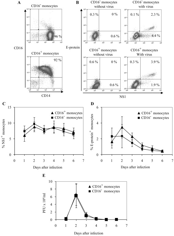
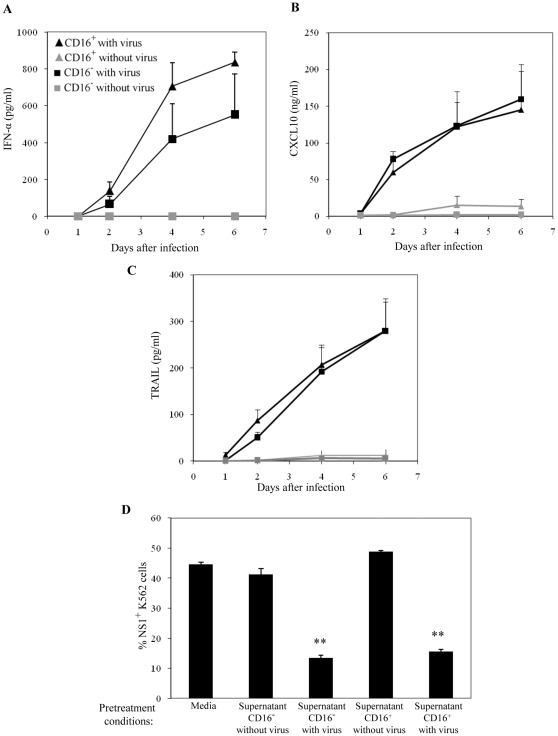
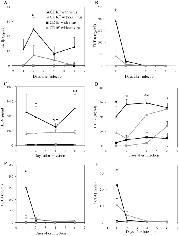
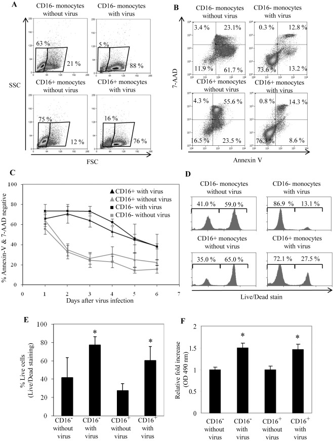
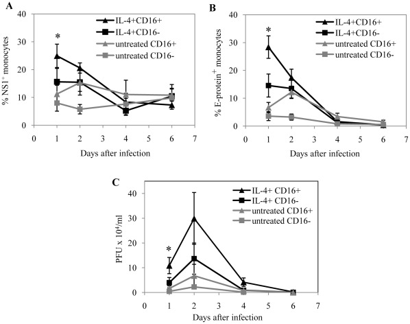
Human blood monocytes play a central role in dengue infections and form the majority of virus infected cells in the blood. Human blood monocytes are heterogeneous and divided into CD16(-) and CD16(+) subsets. Monocyte subsets play distinct roles during disease, but it is not currently known if monocyte subsets differentially contribute to dengue protection and pathogenesis. Here, we compared the susceptibility and response of the human CD16(-) and CD16(+) blood monocyte subsets to primary dengue virus in vitro. We found that both monocyte subsets were equally susceptible to dengue virus (DENV2 NGC), and capable of supporting the initial production of new infective virus particles. Both monocyte subsets produced anti-viral factors, including IFN-α, CXCL10 and TRAIL. However, CD16(+) monocytes were the major producers of inflammatory cytokines and chemokines in response to dengue virus, including IL-1β, TNF-α, IL-6, CCL2, 3 and 4. The susceptibility of both monocyte subsets to infection was increased after IL-4 treatment, but this increase was more profound for the CD16(+) monocyte subset, particularly at early time points after virus exposure. These findings reveal the differential role that monocyte subsets might play during dengue disease.
Conflict of interest statement
Figures
Similar articles
-
Differential regulation of toll-like receptor-2, toll-like receptor-4, CD16 and human leucocyte antigen-DR on peripheral blood monocytes during mild and severe dengue fever.Immunology. 2010 Jun;130(2):202-16. doi: 10.1111/j.1365-2567.2009.03224.x. Epub 2010 Jan 27. Immunology. 2010. PMID: 20113369 Free PMC article.
-
A Novel, Five-Marker Alternative to CD16-CD14 Gating to Identify the Three Human Monocyte Subsets.Front Immunol. 2019 Jul 26;10:1761. doi: 10.3389/fimmu.2019.01761. eCollection 2019. Front Immunol. 2019. PMID: 31402918 Free PMC article.
-
Infliximab therapy increases the frequency of circulating CD16(+) monocytes and modifies macrophage cytokine response to bacterial infection.Clin Exp Immunol. 2014 Sep;177(3):703-11. doi: 10.1111/cei.12375. Clin Exp Immunol. 2014. PMID: 24816497 Free PMC article.
-
Mechanisms of monocyte cell death triggered by dengue virus infection.Apoptosis. 2018 Dec;23(11-12):576-586. doi: 10.1007/s10495-018-1488-1. Apoptosis. 2018. PMID: 30267240 Review.
-
The monocyte-macrophage-mast cell axis in dengue pathogenesis.J Biomed Sci. 2018 Nov 8;25(1):77. doi: 10.1186/s12929-018-0482-9. J Biomed Sci. 2018. PMID: 30409217 Free PMC article. Review.
Cited by
-
Elevated NS1 serum levels reduce CD119 expression and CXCL-10 synthesis in patients with dengue hemorrhagic fever.Rev Soc Bras Med Trop. 2024 Jul 29;57:e00410. doi: 10.1590/0037-8682-0577-2023. eCollection 2024. Rev Soc Bras Med Trop. 2024. PMID: 39082520 Free PMC article.
-
Integrated Transcriptomics Establish Macrophage Polarization Signatures and have Potential Applications for Clinical Health and Disease.Sci Rep. 2015 Aug 25;5:13351. doi: 10.1038/srep13351. Sci Rep. 2015. PMID: 26302899 Free PMC article.
-
Immunomodulation in dengue: towards deciphering dengue severity markers.Cell Commun Signal. 2024 Sep 26;22(1):451. doi: 10.1186/s12964-024-01779-4. Cell Commun Signal. 2024. PMID: 39327552 Free PMC article. Review.
-
Amaryllidaceae Alkaloid Cherylline Inhibits the Replication of Dengue and Zika Viruses.Antimicrob Agents Chemother. 2021 Aug 17;65(9):e0039821. doi: 10.1128/AAC.00398-21. Epub 2021 Aug 17. Antimicrob Agents Chemother. 2021. PMID: 34152811 Free PMC article.
-
Proteome modulation triggers potent antiviral response in Japanese Encephalitis Virus infected human macrophages.Arch Microbiol. 2024 Nov 9;206(12):464. doi: 10.1007/s00203-024-04167-1. Arch Microbiol. 2024. PMID: 39520552
References
-
- Ziegler Heitbrock HW. Heterogeneity of human blood monocytes: the CD14+ CD16+ subpopulation. Immunol. Today. 1996;17:424–428. - PubMed
-
- Passlick B, Flieger D, Ziegler Heitbrock HW. Identification and characterization of a novel monocyte subpopulation in human peripheral blood. Blood. 1989;74:2527–2534. - PubMed
-
- Frankenberger M, Sternsdorf T, Pechumer H, Pforte A, Ziegler Heitbrock HW. Differential cytokine expression in human blood monocyte subpopulations: a polymerase chain reaction analysis. Blood. 1996;87:373–377. - PubMed
-
- Belge KU, Dayyani F, Horelt A, Siedlar M, Frankenberger M, et al. The proinflammatory CD14+CD16+DR++ monocytes are a major source of TNF. J. Immunol. 2002;168:3536–3542. - PubMed
-
- Weber C, Belge KU, von HP, Draude G, Steppich B, et al. Differential chemokine receptor expression and function in human monocyte subpopulations. J. Leukoc. Biol. 2000;67:699–704. - PubMed
Publication types
MeSH terms
Substances
LinkOut - more resources
Full Text Sources
Other Literature Sources
