

Locating the binding sites of Pb(II) ion with human and bovine serum albumins
- PMID: 22574219
- PMCID: PMC3344939
- DOI: 10.1371/journal.pone.0036723
Locating the binding sites of Pb(II) ion with human and bovine serum albumins
Abstract
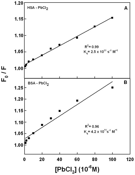
Lead is a potent environmental toxin that has accumulated above its natural level as a result of human activity. Pb cation shows major affinity towards protein complexation and it has been used as modulator of protein-membrane interactions. We located the binding sites of Pb(II) with human serum (HSA) and bovine serum albumins (BSA) at physiological conditions, using constant protein concentration and various Pb contents. FTIR, UV-visible, CD, fluorescence and X-ray photoelectron spectroscopic (XPS) methods were used to analyse Pb binding sites, the binding constant and the effect of metal ion complexation on HSA and BSA stability and conformations. Structural analysis showed that Pb binds strongly to HSA and BSA via hydrophilic contacts with overall binding constants of K(Pb-HSA) = 8.2 (±0.8)×10(4) M(-1) and K(Pb-BSA) = 7.5 (±0.7)×10(4) M(-1). The number of bound Pb cation per protein is 0.7 per HSA and BSA complexes. XPS located the binding sites of Pb cation with protein N and O atoms. Pb complexation alters protein conformation by a major reduction of α-helix from 57% (free HSA) to 48% (metal-complex) and 63% (free BSA) to 52% (metal-complex) inducing a partial protein destabilization.
Conflict of interest statement
Figures


References
-
- http://www.cdc.gov./nceh/lead/, Center for Disease Control and Prevention, Accessed November 1, 2011.
-
- Eslam E, Liu D, Li T, Yang X, Jin X, et al. Efect of Pb toxicity on leaf growth, physiology and ultrastructure in the two ecotypes of Elsholtzia argyi. J Hazard Mater. 2008;154:914–926. - PubMed
-
- Qufei L, Fashui H. Effects of Pb2+ on the structure and function of photosystem II of Spirodela polyrrhiza, Biol Trace Elem Res. 2009;129:251–260. - PubMed
Publication types
MeSH terms
Substances
LinkOut - more resources
Full Text Sources
