. 2012 May 10;12(1):17.
doi: 10.1186/1475-2867-12-17.

Paralemmin-1 is over-expressed in estrogen-receptor positive breast cancers

Affiliations

Paralemmin-1 is over-expressed in estrogen-receptor positive breast cancers

Casey M Turk et al. Cancer Cell Int. .

Abstract

Background: Paralemmin-1 is a phosphoprotein lipid-anchored to the cytoplasmic face of membranes where it functions in membrane dynamics, maintenance of cell shape, and process formation. Expression of paralemmin-1 and its major splice variant (Δ exon 8) as well as the extent of posttranslational modifications are tissue- and development-specific. Paralemmin-1 expression in normal breast and breast cancer tissue has not been described previously.

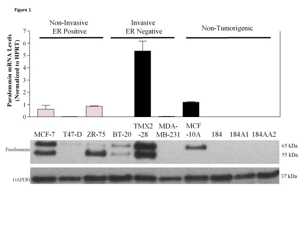
Results: Paralemmin-1 mRNA and protein expression was evaluated in ten breast cell lines, 26 primary tumors, and 10 reduction mammoplasty (RM) tissues using real time RT-PCR. Paralemmin-1 splice variants were assessed in tumor and RM tissues using a series of primers and RT-PCR. Paralemmin-1 protein expression was examined in cell lines using Western Blots and in 31 ductal carcinomas in situ, 65 infiltrating ductal carcinomas, and 40 RM tissues using immunohistochemistry. Paralemmin-1 mRNA levels were higher in breast cancers than in RM tissue and estrogen receptor (ER)-positive tumors had higher transcript levels than ER-negative tumors. The Δ exon 8 splice variant was detected more frequently in tumor than in RM tissues. Protein expression was consistent with mRNA results showing higher paralemmin-1 expression in ER-positive tumors.

Conclusions: The differential expression of paralemmin-1 in a subset of breast cancers suggests the existence of variation in membrane dynamics that may be exploited to improve diagnosis or provide a therapeutic target.


Figures

Figure 1
Figure 1
Paralemmin-1 is differentially expressed in breast epithelial cell lines. RNA and protein lysates were isolated from tumorigenic and non-tumorigenic breast cell lines. Top: Real time qRT-PCR shows mRNA expression of paralemmin-1; means and S.E. from three separate cell cultures are presented; ER-positive cell lines, (hatched bars), ER-negative cell lines (solid bars). Bottom: Protein lysates (15 μg) were probed for paralemmin-1 expression by Western immunoblotting. Image is a representative of at least three separate experiments with different biological samples.
Figure 2
Figure 2
Paralemmin-1 mRNA is highly expressed in ER-positive human tumor samples. Gene expression of paralemmin-1 was determined in 26 frozen breast carcinoma tissue (T) and 10 reduction mammoplasty (RM) samples (open bars) using real time RT-PCR. ER-positive tumors were assigned the numbers 1–18 (red hatched bars) while ER-negative tumors were assigned 19–30 (solid bars) and then sorted by paralemmin-1 expression.
Figure 3
Figure 3
Scoring system used to evaluate paralemmin-1 expression in TMA from breast tumors and reduction mammoplasty specimens. Intensity of immunoreactivity of epithelial cells was used to assign scores of 0, 1, 2, and 3. A 0 was little to no staining, a 1 was considered to be “light” intensity of staining; a 2 was considered to be “moderate” intensity of staining, a 3 was considered to be “strong” intensity of staining. Images are from a DCIS TMA with inset in score 1, 2 and 3 images showing increasing intensity of paralemmin-1 plasma membrane staining in tumor cells. Image for score 0 shows negative paralemmin-1 staining in tumor cells and small peritumoral lymphocytes (left). Intratumoral and peritumoral vascular endothelial cells (*) and elongated stromal cells are positive for paralemmin-1. Scale bars 50 micron.
Figure 4
Figure 4
Paralemmin-1 expression in breast epithelial cells is primarily localized to the cell membrane and is more highly expressed in tumor tissue than reduction mammoplasty. Breast tumor TMAs and reduction mammoplasty tissues were prepared, stained, and scored as described in the Methods. Representative images of paralemmin-1 immunoreactivity in reduction mammoplasty tissues (A and B, low and high magnification) show partial staining in terminal duct and acinar ductal epithelial cells. Paralemmin-1 is present in stromal cells and endothelial cells (image B *). In contrast, in tumor tissues (C and D, high magnification) significant immunoreactivity is present in the malignant epithelial cells where the staining is localized to the cell membrane. Scale bar image A 100 micron. Scale bars image B, C, D 50 micron.
Figure 5
Figure 5
Tumor tissues and breast cell lines express a higher proportion of the Δ exon 8 splice variant of paralemmin-1 than do reduction mammoplasty tissues. RNA was isolated and amplified with RT-PCR analysis using a primer set to detect Δ exon 8 splice variant. RT-PCR products were separated on a 2% low melting agarose gel and visualized by ethidium bromide. The full abbreviations of the cell lines are in Table 1. Tumor tissue samples are represented by the prefix T and reduction mammoplasty tissue samples are represented by the prefix R. Numbers on the left of the figures represent the full length product (275 bp) and the Δ exon 8 splice variant (143 bp).

References

    1. Kutzleb C, Petrasch Parwez E, Kilimann MW. Cellular and subcellular localization of paralemmin-1, a protein involved in cell shape control, in the rat brain, adrenal gland and kidney. Histochem Cell Biol. 2007;127(1):13–30. - PubMed
    1. Kutzleb C, Sanders G, Yamamoto R, Wang X, Lichte B, Petrasch Parwez E, Kilimann MW. Paralemmin, a prenyl-palmitoyl-anchored phosphoprotein abundant in neurons and implicated in plasma membrane dynamics and cell process formation. J Cell Biol. 1998;143(3):795–813. doi: 10.1083/jcb.143.3.795. - DOI - PMC - PubMed
    1. Arstikaitis P, Gauthier Campbell C, Carolina Gutierrez Herrera R, Huang K, Levinson J, Murphy T, Kilimann MW, Sala C, Colicos M, El Husseini A. Paralemmin-1, a modulator of filopodia induction is required for spine maturation. Mol Biol Cell. 2008;19(5):2026–2038. doi: 10.1091/mbc.E07-08-0802. - DOI - PMC - PubMed
    1. Basile M, Lin R, Kabbani N, Karpa K, Kilimann MW, Simpson I, Kester M. Paralemmin interacts with D3 dopamine receptors: implications for membrane localization and cAMP signaling. Arch Biochem Biophys. 2006;446(1):60–68. doi: 10.1016/j.abb.2005.10.027. - DOI - PubMed
    1. Gozgit JM, Pentecost BT, Marconi SA, Otis CN, Wu C, Arcaro KF. Use of an aggressive MCF-7 cell line variant, TMX2-28, to study cell invasion in breast cancer. Mol Cancer Res. 2006;4(12):905–913. doi: 10.1158/1541-7786.MCR-06-0147. - DOI - PubMed

LinkOut - more resources