. 2012 Aug;23(8):891-902.
doi: 10.1089/hum.2012.052.

Safety study of adeno-associated virus serotype 2-mediated human acid sphingomyelinase expression in the nonhuman primate brain

Affiliations

Safety study of adeno-associated virus serotype 2-mediated human acid sphingomyelinase expression in the nonhuman primate brain

Ernesto A Salegio et al. Hum Gene Ther. 2012 Aug.

Abstract

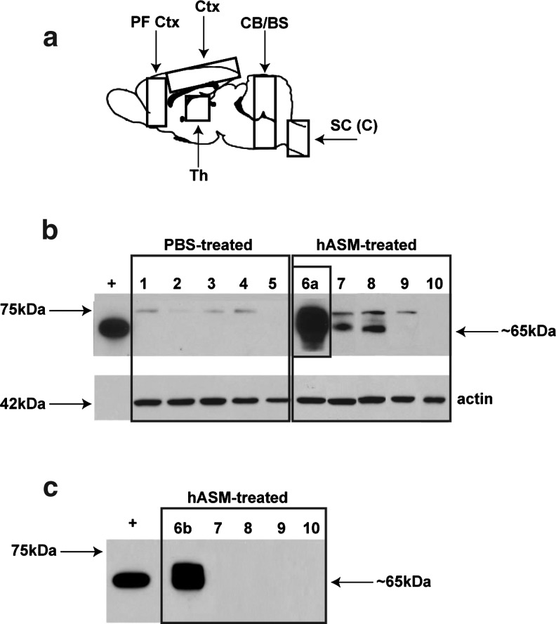
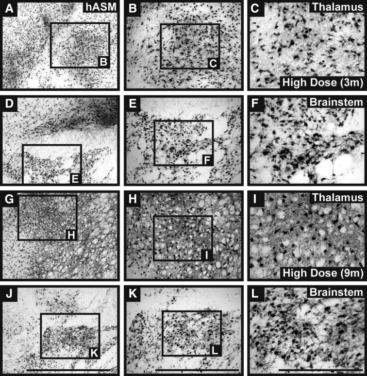
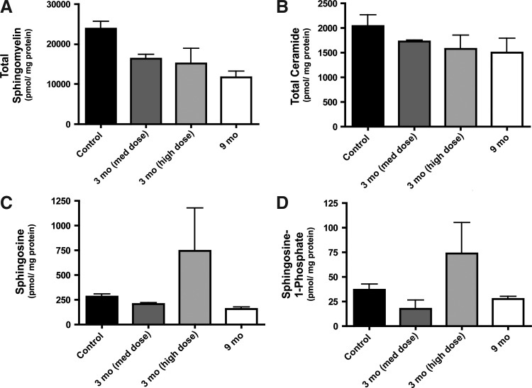
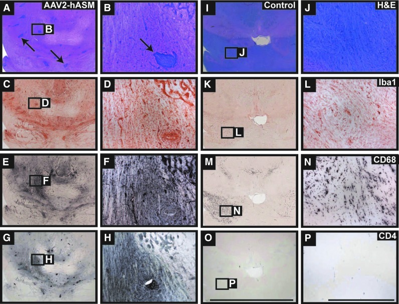
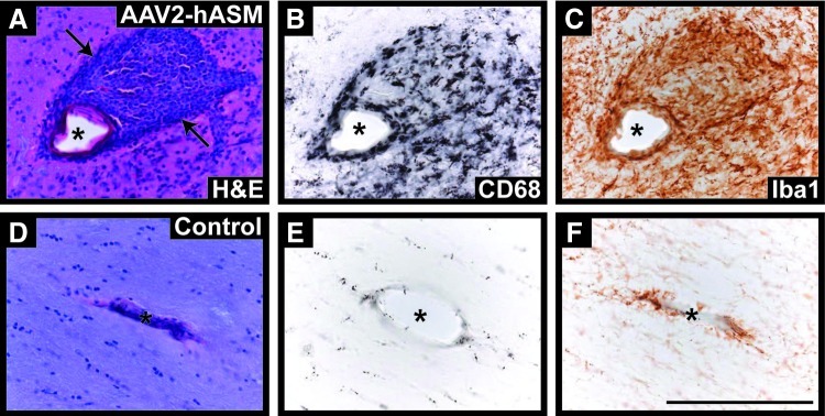
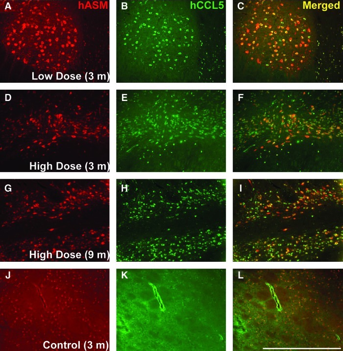
Niemann-Pick disease is a lysosomal storage disorder resulting from inherited deficiency in acid sphingomyelinase (ASM). Use of adeno-associated virus serotype 2 (AAV2) to deliver human acid sphingomyelinase (hASM) is currently being explored as a means to treat the devastating neurological features of NPD, which are refractory to traditional enzyme replacement therapy. In this study, we evaluated the long-term efficacy and safety of AAV2-hASM after direct infusion into the CNS of nonhuman primates. First, we confirmed the efficacy of AAV2-hASM in naive rats, which exhibited increased ASM expression and enzyme activity after infusion, without evidence of local or systemic toxicity. Next, the model was adapted to naive nonhuman primates (NHPs) with various doses of AAV2-hASM or saline delivered into the brainstem and both thalami. Strikingly, NHPs that received a high dose of AAV2-hASM displayed significant motor deficits that were not seen in low-dose animals in both the short-term (3-month) and long-term (9-month) treatment groups. In treated NHPs, ASM expression and activity were elevated with associated alterations in the sphingolipidomic profile in brain regions transduced with AAV2-hASM. Initial histological analysis indicated marked inflammatory reactions, and immunohistochemical analysis confirmed a robust inflammatory response. Importantly, pronounced upregulation of the chemokine CCL5, a target of ASM-mediated inflammatory signaling, was detected that correlated with the inflammatory response, providing a possible mechanism for hASM-associated toxicity. This study defines dose-dependent and dose-independent toxicities of AAV2-hASM in the naive primate brain, and reveals potential challenges in the design of a clinical trial.


Figures

FIG. 1.
FIG. 1.
Western blot analysis of tissue lysates from rats. (a) Schematic of the rat brain, indicating dissected regions used for Western blot analysis. CB/BS, cerebellum/brainstem; Ctx, cortex; PF Ctx, prefrontal cortex; SC (C), spinal cord cervical region; Th, thalamus. (b) Analysis of long-exposure Western blots indicated the presence of two proteins: (1) a 75-kDa band believed to represent endogenous acid sphingomyelinase (ASM), found in all samples except those collected from upper spinal cord regions, and (2) a 65-kDa band indicative of the mature enzyme and of similar molecular weight to the positive control (partially purified lysosomal hASM) and detected only in vector-treated samples from the thalamus, cortex, and prefrontal cortex. (c) In the shorter exposure Western blot, bands corresponding to the endogenous protein were not detected in any samples. Only samples collected from thalamic regions demonstrated the presence of a 65-kDa protein. Thalamus (lanes 1, 6a, and 6b), cortex (lanes 2 and 7), prefrontal cortex (lanes 3 and 8), cerebellum/brainstem (lanes 4 and 9), cervical/thoracic spinal cord (lanes 5 and 10), partially purified lysosomal hASM (+; positive control), and β-actin (loading control).
FIG. 2.
FIG. 2.
Parenchymal neuronal ASM expression in nonhuman primates (NHPs). Immunostaining against hASM indicated strong neuronal transduction in both targeted regions (thalamus and brainstem). Expression of the hASM transgene was detected in all vector-treated brains irrespective of AAV dose and survival time point. No hASM+ cells were found in the brains of control animals (data not shown). Scale bars: (A, D, G, and J) 2 mm; (B, E, H, and K) 1 mm; (C, F, I, and L) 500 μm.
FIG. 3.
FIG. 3.
Lipid analysis of NHP brain samples. Thalamic homogenates from PBS-treated controls and AAV2-hASM-treated NHPs were submitted for sphingolipidomic analysis, as described in Materials and Methods. Data are expressed as picomoles per milligram of total protein for each lipid or group of lipids analyzed (n=2 per group). (A) Total sphingomyelin; (B) total ceramide; (C) sphingosine; (D) sphingosine 1-phosphate.
FIG. 4.
FIG. 4.
Brain pathology and inflammatory cell recruitment. Histological examination of vector-treated NHP brains demonstrated significant pathological signs (A and B, arrows) not observed in PBS-treated controls. Immunostaining of thalamic and brainstem regions in hASM-infused animals indicated robust infiltration and activation of inflammatory cells consisting mainly of microglia (C and D, Iba1+), macrophages (E and F, CD68+), and activated T cells (G and H, CD4+). Expression of inflammatory cells in control animals represents the normal immune response to cannula penetration and the convection-enhanced delivery (CED) procedure within the parenchyma (I–P). Scale bars: Low-magnification panels, 10 mm; high-magnification panels, 1 mm. Color images available online at www.liebertpub.com/hum
FIG. 5.
FIG. 5.
Perivascular cuffing in AAV2-hASM-treated NHP brains. (A) Hematoxylin and eosin-processed sections from vector-treated animals (high dose, 3-month survival) indicate the presence of cuffing (arrows) around blood vessels (asterisks); cuffs were composed of macrophage (CD68+) and microglial (Iba1+) infiltrates (B and C), as compared with PBS-treated control tissue (D–F). Scale bar: 200 μm. Color images available online at www.liebertpub.com/hum
FIG. 6.
FIG. 6.
Neuronal coexpression of hASM and CCL5. Immunofluorescence staining for hASM (red) and the chemokine CCL5/RANTES (green) revealed neuronal coexpression of hASM and CCL5 in vector-treated animals independent of AAV dose and survival (A–I, yellow), compared with PBS-treated controls (J–L). Note that neurons staining for both hASM and CCL5 were found only within the brains of NHPs infused with AAV2-hASM. Scale bar: (A–L) 1 mm. Color images available online at www.liebertpub.com/hum

References

    1. Amalfitano A. McVie-Wylie A.J. Hu H., et al. Systemic correction of the muscle disorder glycogen storage disease type II after hepatic targeting of a modified adenovirus vector encoding human acid-α-glucosidase. Proc. Natl. Acad. Sci. U.S.A. 1999;96:8861–8866. - PMC - PubMed
    1. Ayuso E. Mingozzi F. Montane J., et al. High AAV vector purity results in serotype- and tissue-independent enhancement of transduction efficiency. Gene Ther. 2010;17:503–510. - PubMed
    1. Barranger J.M. Novelli E.A. Gene therapy for lysosomal storage disorders. Expert Opin. Biol. Ther. 2001;1:857–867. - PubMed
    1. Bielawski J. Szulc Z.M. Hannun Y.A., et al. Simultaneous quantitative analysis of bioactive sphingolipids by high-performance liquid chromatography-tandem mass spectrometry. Methods. 2006;39:82–91. - PubMed
    1. Bosch A. Perret E. Desmaris N., et al. Reversal of pathology in the entire brain of mucopolysaccharidosis type VII mice after lentivirus-mediated gene transfer. Hum. Gene Ther. 2000;11:1139–1150. - PubMed

Publication types

Substances