. 2012 Aug;236(2):351-62.
doi: 10.1016/j.expneurol.2012.04.018. Epub 2012 May 1.

Macrophage migration inhibitory factor (MIF) is essential for inflammatory and neuropathic pain and enhances pain in response to stress

Affiliations

Macrophage migration inhibitory factor (MIF) is essential for inflammatory and neuropathic pain and enhances pain in response to stress

Jessica K Alexander et al. Exp Neurol. 2012 Aug.

Abstract

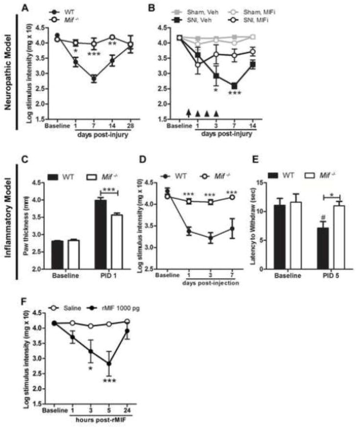
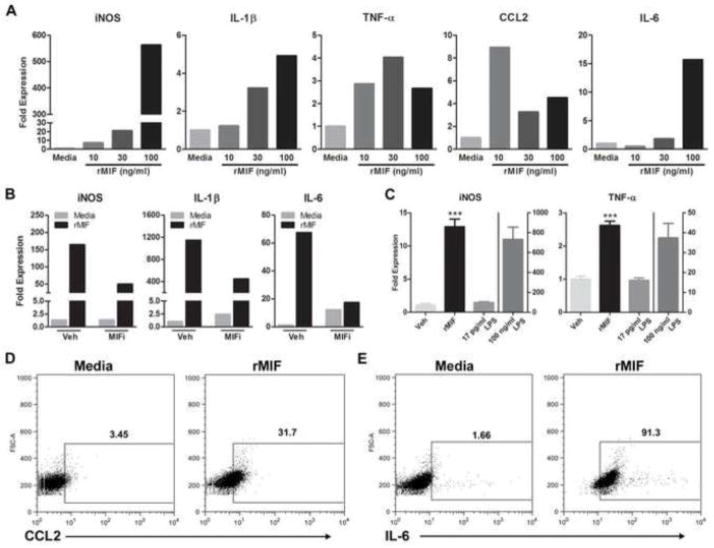
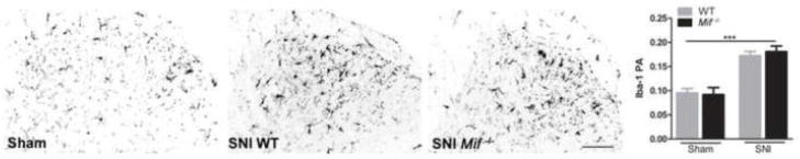
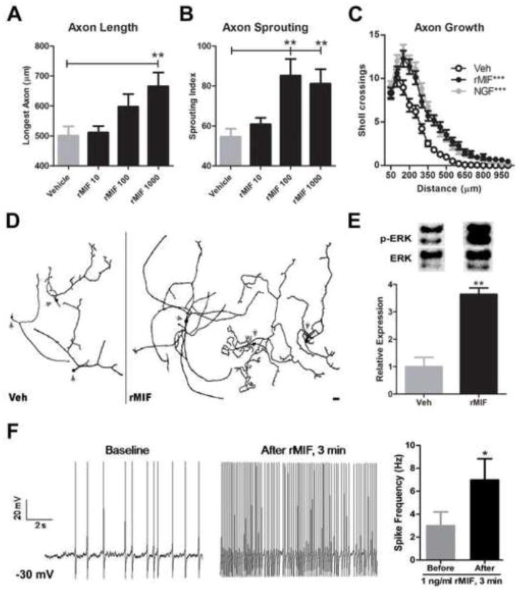
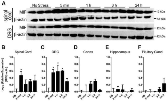
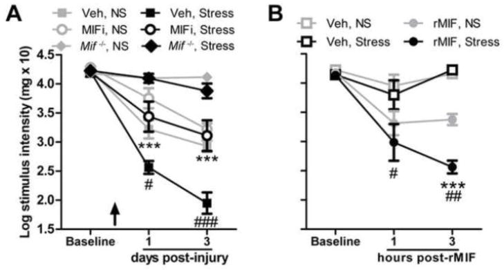
Stress and glucocorticoids exacerbate pain via undefined mechanisms. Macrophage migration inhibitory factor (MIF) is a constitutively expressed protein that is secreted to maintain immune function when glucocorticoids are elevated by trauma or stress. Here we show that MIF is essential for the development of neuropathic and inflammatory pain, and for stress-induced enhancement of neuropathic pain. Mif null mutant mice fail to develop pain-like behaviors in response to inflammatory stimuli or nerve injury. Pharmacological inhibition of MIF attenuates pain-like behaviors caused by nerve injury and prevents sensitization of these behaviors by stress. Conversely, injection of recombinant MIF into naïve mice produces dose-dependent mechanical sensitivity that is exacerbated by stress. MIF elicits pro-inflammatory signaling in microglia and activates sensory neurons, mechanisms that underlie pain. These data implicate MIF as a key regulator of pain and provide a mechanism whereby stressors exacerbate pain. MIF inhibitors warrant clinical investigation for the treatment of chronic pain.


Figures

Figure 1
Figure 1
All offspring from a colony of Mif−/− mice were genotyped via PCR to confirm deletion of Mif. The presence of the neo cassette indicates gene deletion (representative screen from a Mif−/− mouse generated at OSU). See Methods for primers used to amplify Mif and neo cassette.
Figure 2
Figure 2
MIF regulates the onset and magnitude of neuropathic or inflammatory pain-like behaviors. A, Unlike wild type (WT) mice, Mif−/− mice do not develop mechanical hypersensitivity after nerve injury. B, Systemic injection of a MIF inhibitor (MIFi) after nerve injury reduces hypersensitivity (treatments indicated by arrow (60 min post-SNI) and arrowheads (days 1–3 post-SNI)). C, Edema caused by i.pl. CFA is reduced in Mif−/− mice at post-injection day (PID) 1. D, CFA induces mechanical hypersensitivity and E, thermal hyperalgesia in WT mice but not Mif−/− mice. F, Intraplantar injection of rMIF (1000 pg) produces mechanical hypersensitivity. A, B, D, F: y-axis values represent von Frey hair handle markings where 4.31=2 grams of force and 2.83=0.07 grams of force. N=5/group. Results are expressed as mean ± SEM. Statistical comparisons are between Mif−/− and WT mice (A, C, D, E), or SNI, Veh and SNI, MIFi (B) or Saline and rMIF (F) groups., *p<0.05; **p<0.01; ***p<0.0001. In E, # denotes p<0.05 when comparing WT Baseline vs. PID 5.
Figure 3
Figure 3
MIF is constitutively expressed in the rat spinal cord (A–F) and rat peripheral nervous system (G–I). Superimposed confocal images showing MIF (red) and cell-specific markers (green or blue). A,B MIF is expressed in a subset of spinal cord dorsal horn (A) neurons (green; NeuN) but not ventral horn motor neurons (B). C–F, MIF is also expressed in spinal cord axons (C, green: neurofilament (NF), astrocytes (D, green: GFAP), microglia (E, green: OX-42) and oligodendrocytes (F, blue: CC1). G–I, In peripheral nervous system, MIF is expressed in DRG neurons (G, green: NF) and sciatic nerve axons (H, green: NF) but not the myelin sheath (I, green: myelin basic protein (MBP)). J, After nerve injury (12 h post-injury), MIF is expressed by leukocytes, primarily macrophages (green, OX-42) at the epineurium-nerve interface (demarcated by arrows). In the DRG (G), MIF is also apparent in satellite cells (NF-negative cells cuffing neurons, demarcated by arrows). A–J: Scale bars = 20 μm. K, L, Specificity of anti-MIF labeling was confirmed in Mif−/− mouse spinal cord tissue. Despite high background in wild type C57BL/6 mouse tissue, positive labeling is discernible and shown here in dorsal horn cells, including neurons (arrowheads) (K). This labeling is absent in Mif−/− tissue (L). Scale bar for K, L=50 μm.
Figure 4
Figure 4
MIF elicits inflammatory signaling in microglia. A, Treatment of rat primary microglia with rMIF (10, 30 or 100 ng/ml) for 6 h increases iNOS, IL-1β, TNF-α, CCL2, and IL-6 mRNA expression. Data are normalized to an internal control gene (18s) then are expressed relative to mRNA changes elicited by media. Data are representative of three independent experiments. B&C, Specificity of rMIF effects on microglia phenotype. B, Co-application of MIF inhibitor (MIFi; 100 μM) with rMIF for 6 h reduces rMIF-induced cytokine gene expression. C, rMIF effects on microglia phenotype are not due to endotoxin contamination - only trace amounts (17 pg/ml) of endotoxin were detected in rMIF. When treated with 17 pg/ml LPS for 6 h, BV-2 microglia did not increase expression of iNOS or TNF-α. Conversely, 6 h of stimulation with rMIF (100 ng/ml) or high concentrations of LPS (100 ng/ml) markedly induced expression of cytokines (***p < 0.0001 vs. Veh and 17 pg/ml LPS). D&E, rMIF treatment of BV-2 microglia for 12h increases CCL2 and IL-6 protein. Representative contour plots show relative expression profiles for D, CCL2 and E, IL-6 in CD11b+ BV-2 microglia after 12 h of stimulation with media or rMIF (100 ng/ml). The mean percent of CCL2 or IL-6 expressing CD11b+ cells is indicated for each group. Dot plots are representative of two independent replicate experiments. Results are expressed as mean ± SEM.
Figure 5
Figure 5
Spinal cord dorsal horn microglial activation ipsilateral to nerve injury (SNI). Iba-1 immunoreactivity is increased by nerve injury compared to sham at 3 days (* vs. sham of same genotype). The microglial response to injury (or sham) does not differ between WT and Mif −/− mice. N=3 sham; N=5 SNI. PA: proportional area. Scale bar = 100 μm. Results are expressed as mean ± SEM. ***p<0.0001
Figure 6
Figure 6
rMIF increases structural and functional plasticity in sensory neurons. Twenty-four hours after plating, adult DRG neurons were treated with rMIF (10, 100, 1000 pg/ml). Forty eight hours later, the overall length and branching complexity of DRG axons was examined by Sholl analysis. A, rMIF (1000 pg/ml) increased long-distance axon growth and B, sprouting/branching complexity. C, A composite view of axon length (distance) and sprouting shows that rMIF and NGF have nearly identical effects on promoting DRG axon growth (***: vs. Veh; p>0.05 rMIF vs. NGF.) D, Representative DRG neurons (arrows delineate somata) treated with vehicle (Veh) or rMIF (1000 pg/ml) illustrate the neurotrophic effects of rMIF. Data in A–D obtained from n= 75–100 neurons per group (see Methods). Scale bar in D = 100μm. E, ERK activation in DRG neurons after 10 min Veh or rMIF (1000 pg/ml) treatment. F, MIF increases the excitability of small diameter DRG neurons. Current-clamp recordings from the same neuron showing the effects of exposure to rMIF (1 ng/ml). Mean neuron spike frequency measured after current clamp at −30 mV and after 3 min rMIF exposure (n=7 neurons). Results are expressed as mean ± SEM. Unless noted otherwise above, all statistical comparisons were made between rMIF vs. Veh treatments; *p<0.05; **p<0.01; ***p<0.0001.
Figure 7
Figure 7
Tissue MIF levels are increased by acute stress. A, Western blot analysis of MIF in spinal cord and DRG after stress. B–F, Quantitation of stress-induced MIF expression. Tissue MIF levels were normalized to β-actin and expressed relative to No Stress samples (NS) then log transformed to normalize distribution. Samples were collected 5 min or 1, 3 or 24 h after the onset of restraint stress or no stress. N=3 mice/per time point. Data expressed as mean ± SEM. *p<0.05
Figure 8
Figure 8
MIF inhibition prevents stress-enhanced mechanical hypersensitivity. A, A single systemic injection of MIFi prior to stress (demarcated by arrow) prevents stress-enhanced hypersensitivity caused by nerve injury. (#: Veh, No Stress (NS) vs. Veh, Stress; *: Veh, Stress vs. MIFi, Stress). Pain-like responses cannot be elicited by SNI nor are they enhanced by stress in Mif−/− mice. B, Acute stress prior to i.pl. rMIF (1000 pg) increases mechanical hypersensitivity (* vs. rMIF, No Stress (NS); #: Veh, NS vs. rMIF, NS). y-axis values represent von Frey hair handle markings where 4.31=2 grams of force and 2.83=0.07 grams of force. N=5/group. Results are expressed as mean ± SEM. #/*p<0.05; ##/**p<0.01; ###/***p<0.0001

References

    1. Abbadie C, Lindia JA, Cumiskey AM, Peterson LB, Mudgett JS, Bayne EK, DeMartino JA, MacIntyre DE, Forrest MJ. Impaired neuropathic pain responses in mice lacking the chemokine receptor CCR2. Proc Natl Acad Sci U S A. 2003;100:7947–7952. - PMC - PubMed
    1. Alexander JK, DeVries AC, Kigerl KA, Dahlman JM, Popovich PG. Stress exacerbates neuropathic pain via glucocorticoid and NMDA receptor activation. Brain Behav Immun. 2009;23:851–860. - PMC - PubMed
    1. Almad A, McTigue DM. Chronic expression of PPAR-delta by oligodendrocyte lineage cells in the injured rat spinal cord. J Comp Neurol. 2010;518:785–799. - PMC - PubMed
    1. Aloisi AM, Pari G, Ceccarelli I, Vecchi I, Ietta F, Lodi L, Paulesu L. Gender-related effects of chronic non-malignant pain and opioid therapy on plasma levels of macrophage migration inhibitory factor (MIF) Pain. 2005;115:142–151. - PubMed
    1. Ankeny DP, Guan Z, Popovich PG. B cells produce pathogenic antibodies and impair recovery after spinal cord injury in mice. J Clin Invest. 2009;119:2990–2999. - PMC - PubMed

Publication types

MeSH terms

Substances