. 2012 May;98(1-2):23-30.
doi: 10.1016/j.prostaglandins.2012.03.002. Epub 2012 Apr 2.

E-prostanoid 2 receptor signaling suppresses lung innate immunity against Streptococcus pneumoniae

Affiliations

E-prostanoid 2 receptor signaling suppresses lung innate immunity against Streptococcus pneumoniae

David M Aronoff et al. Prostaglandins Other Lipid Mediat. 2012 May.

Abstract

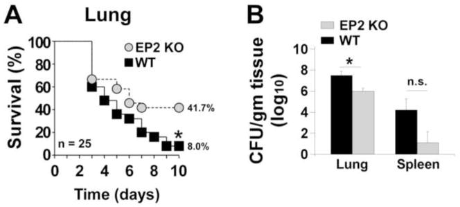
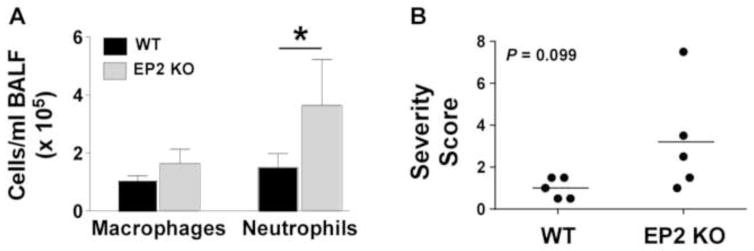
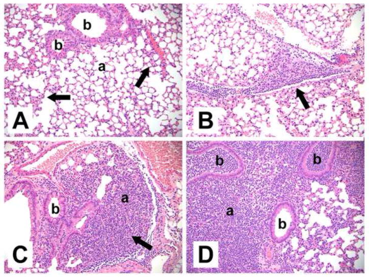
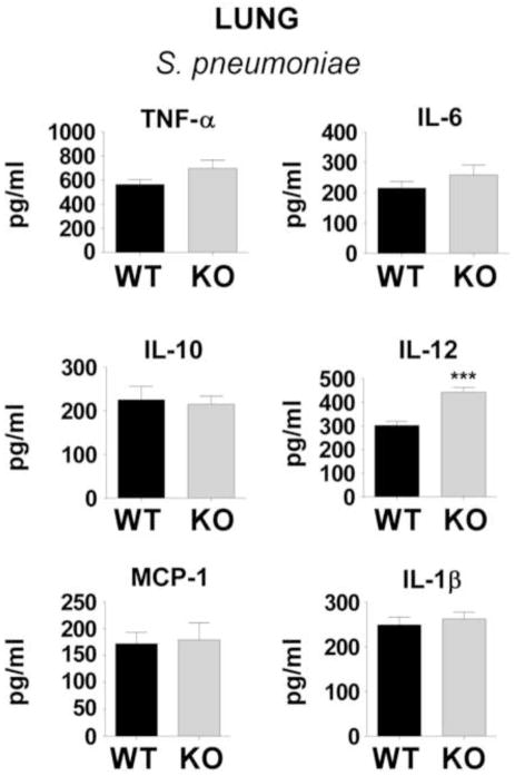
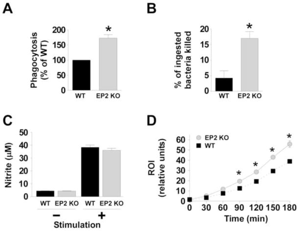
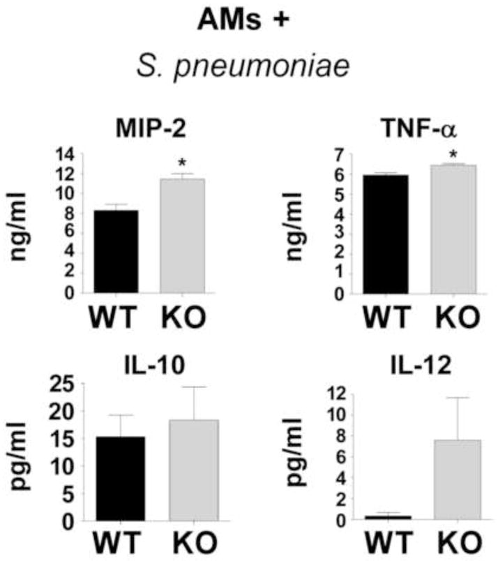
Pneumonia is a major global health problem. Prostaglandin (PG) E(2) is an immunomodulatory lipid with anti-inflammatory, immunosuppressive, and pro-resolving actions. Data suggest that the E-prostanoid (EP) 2 receptor mediates immunomodulatory effects of PGE(2), but the extent to which this occurs in Streptococcus pneumoniae infection is unknown. Intratracheal lung infection of C57BL/6 mice possessing (EP2(+/+)) or lacking (EP2(-/-)) the EP2 receptor was performed, as were in vitro studies of alveolar macrophage (AM) host defense functions. Bacterial clearance and survival were significantly improved in vivo in EP2(-/-) mice and it correlated with greater neutrophilic inflammation and higher lung IL-12 levels. Upon ex vivo challenge with pneumococcus, EP2(-/-)cells expressed greater amounts of TNF-α and MIP-2 than did EP2(+/+) AMs, and had improved phagocytosis, intracellular killing, and reactive oxygen intermediate generation. These data suggest that PGE(2)-EP2 signaling may provide a novel pharmacological target for treating pneumococcal pneumonia in combination with antimicrobials.


Conflict of interest statement

All the authors declare that they have no competing interests.

Figures

Fig. 1
Fig. 1. Improved survival and enhanced bacterial containment after pneumococcal infection in mice lacking the EP2 receptor
Female wild type (WT) C57BL/6 or EP2 receptor knockout (EP2 KO) mice were infected intratracheally (A) with S. pneumoniae as detailed in the Materials and Methods section. Survival was monitored for 10 days. The number of mice per group is shown. *P < 0.05 by Mantel-Cox log-rank test. (B) Bacterial loads (CFU S. pneumoniae per gram of homogenized tissue) in the lung and spleen of WT and EP2 KO mice 24 h after infection (n = 5 mice per experimental group). *P < 0.05 by Student t-test; n.s., not significant. Data from one representative experiment of two independent experiments yielding similar results are shown.
Fig. 2
Fig. 2. Enhanced acute inflammation in EP2 receptor deficient mice after S. pneumoniae lung infection
(A) Bronchoalveolar lavage fluid (BALF) cell counts were determined in wild type (WT) and EP2 knockout (KO) mice as detailed in the Materials and Methods section 24 h after intratracheal inoculation with S. pneumoniae. *P < 0.05 by Student t-test (n = 5 mice per group). (B) Pulmonary inflammation was quantitated using a summary severity score calculated through blinded histopathological analysis as described in the Materials and Methods section. EP2 KO mice demonstrated a trend towards more severe acute inflammation in the lungs 24 hr after infection. *P = 0.099 by Student t-test. Data from one representative experiment of two independent experiments yielding similar results are shown
Fig. 3
Fig. 3. Representative histopathological changes in the lungs of S. pneumoniae-infected wild type (WT) and EP2 knockout (KO) mice 24 h after inoculation
Abbreviations: b: bronchiole, a: alveoli. (A) Infected WT mouse without significant inflammation (summary score 0.5). The majority of alveolar airspaces are clear. There are small areas of increased interstitial macrophagic cellularity (arrows). (B) Infected WT mouse with mild, focal neutrophilic inflammation (summary score 1.0), adjacent to a pulmonary blood vessel and along interlobular septae (arrow). (C) Infected EP2−/− mouse with mild-moderate inflammation (summary score 1.5) focally obscuring the alveoli (arrow). (D) Infected EP2−/− mouse with focally intense inflammation (summary score 7.5) consisting of neutrophils extensively filling alveoli and bronchioles, pleural involvement (not shown), and increased interstitial cellularity. Hematoxylin and eosin. Original magnifications x200.
Fig. 4
Fig. 4. Inflammatory mediators in whole lung homogenates of wild type (WT) C57BL/6 mice or EP2 knockout (KO) mice 24 h after inoculation
Cytokines and chemokines were measured by ELISA as detailed in the Materials and Methods section 24 h after intratracheal inoculation with S. pneumoniae. ***P < 0.001 by Student t-test (n = 5 mice per group). IL, interleukin, MCP, monocyte chemotactic protein-1.
Fig. 5
Fig. 5. Enhanced in vitro alveolar macrophage defense functions in EP2 knockout (KO) mice compared with wild type (WT) alveolar macrophages
Alveolar macrophages were obtained from uninfected WT and EP2 KO mice as described in the Materials and Methods section. (A) Cells were inoculated with heat-inactivated, FITC-labeled S. pneumoniae (multiple of infection 150 bacteria per macrophage) and phagocytosis was measured after 3 hrs. *P < 0.05 by Student t-test. Data represent the mean ± SEM of a minimum of 3 independent experiments performed in octuplet. (B) The survival of internalized S. pneumoniae was determined as noted in the Materials and Methods section. *P < 0.05 by Student t-test. Data represent the mean ± SEM of a minimum of 3 independent experiments performed in triplicate. (C) The capacity for alveolar macrophages to generate nitric oxide (measured as nitrite) was assessed for WT and EP2 KO cells following stimulation with or without 10 μg/ml of lipoteichoic acid and 10 ng/ml IFN-γ for 24h. (D) Alveolar macrophages from WT mice (black squares and dotted line) or EP2 KO mice (grey circles and solid line) were cultured with 2′,7′-dichlorodihydrofluorescein diacetate (H2DCF) for 1h then stimulated with heat-killed S. pneumoniae using a multiplicity of infection of 50:1. Reactive oxygen intermediate (ROI) production was assessed fluorometrically and expressed as relative fluorescence units. The data represent the mean of 3 experiments completed in quadruplicate for each time point. *P < 0.05 by Student t-test.
Fig. 6
Fig. 6. Alveolar macrophage cytokine and chemokine production following stimulation with S. pneumoniae
Alveolar macrophages were obtained from uninfected WT and EP2 knockout (KO) mice and inoculated in vitro with S. pneumoniae as described in the Materials and Methods section. After 24 hrs of inoculation, bacteria- and cell-free supernatants were assayed for inflammatory mediators by ELISA. *P < 0.05 by Student t-test. Data represent the mean ± SEM of n = 3 independent experiments.

References

    1. Bernstein A, Bilheimer LT, Makuc DM. National Center for Health, S. Dept. of Health and Human Services, Centers for Disease Control and Prevention, National Center for Health Statistics. Hyattsville, MD: 2011.
    1. Bosso JA, Drew RH. Application of antimicrobial stewardship to optimise management of community acquired pneumonia. Int J Clin Pract. 2011;65(7):775–83. - PubMed
    1. Mandell LA, Wunderink RG, Anzueto A, et al. Infectious Diseases Society of America/American Thoracic Society consensus guidelines on the management of community-acquired pneumonia in adults. Clin Infect Dis. 2007;44 (Suppl 2):S27–72. - PMC - PubMed
    1. Levine OS, O’Brien KL, Knoll M, et al. Pneumococcal vaccination in developing countries. Lancet. 2006;367(9526):1880–2. - PubMed
    1. Kadioglu A, Andrew PW. The innate immune response to pneumococcal lung infection: the untold story. Trends Immunol. 2004;25(3):143–9. - PubMed

Publication types

MeSH terms

LinkOut - more resources