Review
. 2012 Aug 14;586(17):2638-47.
doi: 10.1016/j.febslet.2012.04.015. Epub 2012 Apr 21.

Plasticity of PDZ domains in ligand recognition and signaling

Affiliations
Review

Plasticity of PDZ domains in ligand recognition and signaling

Ylva Ivarsson. FEBS Lett. .

Abstract

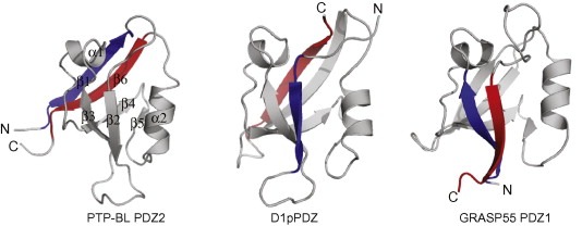
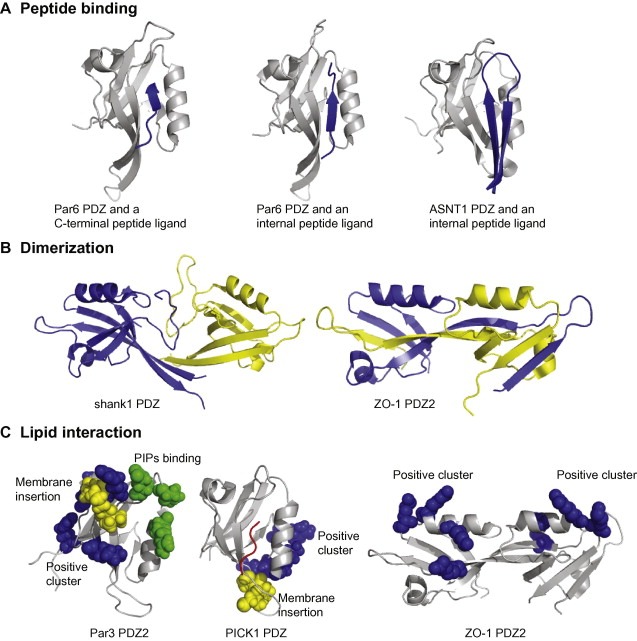
The PDZ domain is a protein-protein interacting module that plays an important role in the organization of signaling complexes. The recognition of short intrinsically disordered C-terminal peptide motifs is the archetypical PDZ function, but the functional repertoire of this versatile module also includes recognition of internal peptide sequences, dimerization and phospholipid binding. The PDZ function can be tuned by various means such as allosteric effects, changes of physiological buffer conditions and phosphorylation of PDZ domains and/or ligands, which poses PDZ domains as dynamic regulators of cell signaling. This review is focused on the plasticity of the PDZ interactions.


Figures

Figure 1
Figure 1
Cartoon representations of canonical and PDZ‐like domains with the N‐ and C‐terminal strands highlighted in blue and red, respectively. PTP‐BL PDZ2 (PDB code http://1GM1, left) with indicated secondary elements (β1–β2–β3–α1–β4–β5–α2–β6) represents a canonical metazoan PDZ domain. The PDZ domain of the D1 C‐terminal‐processing protease (middle, PDB code http://1FC7) and GRASP55 PDZ1 (right, PDB code http://3RLE) illustrate two differentially circularly permuted PDZ structures.
Figure 2
Figure 2
The repertoire of PDZ interactions include peptide binding (A), dimerization (B) and phospholipid interactions (C). (A) Par6 PDZ in complex with a VKESLV‐COO‐ peptide (left; PDB code http://1RZX) representing canonical C‐terminal peptide binding. The structure can be compared with the same protein in complex with a PALS1 internal peptide ligand (middle, PDB code http://1X8S). Note the changes in the β1–β2 loop that allow for the accommodation of the internal peptide ligand. The structure of SNTA1 PDZ in complex with the nNOS internal peptide (right, PDB code http://1QAV) illustrates that internal peptide ligands can be accommodated if they take restricted conformations mimicking free C‐terminal ligands. (B) Different strategies for PDZ–PDZ dimerization as illustrated by the structures of shank1 PDZ (left, PDB code http://1Q3P) and ZO‐1 PDZ2 (right, PDB code http://2RCZ). The constituting monomers are shown in in blue and yellow cartoon representation. (C) PDZ domains employ different means for interacting with PIPs containing lipid membranes. Par3 PDZ2 (PDB code http://2OGP) interacts with negatively charged lipid membranes through a combination of non‐specific electrostatic interactions (Lys491, Arg496, Lys506 and Arg546 indicated in blue spheres), membrane penetration (Leu494, Pro495 and Ile500 in yellow spheres), and a defined PIPs binding site (Glu469, Lys535 and Arg532 in green spheres) [17]. PICK1 PDZ (middle, PDB code http://2PKU) interacts with lipid membranes through a conserved Cys‐Pro‐Cys motif (yellow spheres) and a positive charge cluster (Arg76, Lys79 and Lys81 in blue spheres). A bound peptide is indicated in red. Mutaganic analysis of ZO‐1 PDZ2 (right) indicated a set of basic residues to be of importance for the interactions with PIPs containing liposomes (Arg201, Lys246, Arg251 and Lys253 shown as blue spheres) [86].

References

    1. Good M.C., Zalatan J.G., Lim W.A., Scaffold proteins: hubs for controlling the flow of cellular information. Science, 332, (2011), 680– 686. - PMC - PubMed
    1. Cho K.O., Hunt C.A., Kennedy M.B., The rat brain postsynaptic density fraction contains a homolog of the Drosophila discs-large tumor suppressor protein. Neuron, 9, (1992), 929– 942. - PubMed
    1. Woods D.F., Bryant P.J., ZO-1, DlgA and PSD-95/SAP90: homologous proteins in tight, septate and synaptic cell junctions. Mech. Dev., 44, (1993), 85– 89. - PubMed
    1. Kim E., Niethammer M., Rothschild A., Jan Y.N., Sheng M., Clustering of Shaker-type K+ channels by interaction with a family of membrane-associated guanylate kinases. Nature, 378, (1995), 85– 88. - PubMed
    1. Ponting C.P., Evidence for PDZ domains in bacteria, yeast, and plants. Protein Sci., 6, (1997), 464– 468. - PMC - PubMed