. 2012 Jun;35(5):400-8.
doi: 10.1097/CJI.0b013e31825898c5.

Tumor-specific CD4+ melanoma tumor-infiltrating lymphocytes

Affiliations

Tumor-specific CD4+ melanoma tumor-infiltrating lymphocytes

Kevin M Friedman et al. J Immunother. 2012 Jun.

Abstract

Adoptive cell therapy using tumor-infiltrating lymphocytes (TIL) can mediate objective and durable tumor regressions in patients with metastatic melanoma. CD8+ tumor-reactive TIL are well studied in humans and animals, yet the function of tumor-infiltrating CD4+ T lymphocytes in patient treatments remains controversial. We recently demonstrated that CD4+ TILs are not necessary for objective responses in patients. Coinfusion with tumor-specific CD4 TIL may enhance or increase the durability of tumor regressions, but the number of patients with tumor-reactive CD4 TIL is unknown. We screened 44 CD8+-depleted TIL for in vitro reactivity against autologous tumor. Nine (20%) showed specific reactivity by interferon-γ release assay, of which 8 were specifically blocked by an anti-HLA-DR antibody. Flow-cytometric analysis of these reactive TIL confirmed a high CD4+ composition (median 89%). Highlighting the contribution of CD4+ TIL to tumor regression, a patient with widespread metastatic disease was administered TIL containing HLA class II-restricted tumor activity with high-dose interleukin-2 therapy after lymphodepletion that mediated regression of extensive metastatic disease in the liver and spleen. These results demonstrate that at least 20% of metastatic melanomas contain CD4+ lymphocytes with specific tumor recognition and suggest a possible role for CD4+ cells in the effectiveness of adoptive cell therapy.


Figures

Figure 1
Figure 1. CD4+ Tumor infiltrating lymphocytes (TIL) originate from a resected metastatic lesion via CD8+ depletion and in vitro culture
Tumor specimens were obtained and processed under sterile conditions to single cell suspensions by enzymatic digestion. The first priority for tissue use was to generate a TIL culture for protocol treat; when available, some of the autologous fresh tumor (FrTu) single cell suspension was cryopreserved immediately for use later as “targets” in coculture assays. The single cell suspension was plated in lymphocyte growth media and maintained in vitro until a “bulk” young tumor infiltrating lymphocyte cultures (bulk TIL) was established. The majority of the bulk TIL underwent CD8+ enrichment using a CliniMACS(R) magnetic cell separator. All three fractions, the bulk TIL, the CD8+ enriched fraction, and the CD8+-depleted fraction (containing CD4+ cells) were rapidly expanded with OKT3 (anti-CD3), IL-2 and feeder cells. The resulting TIL cultures were evaluated for phenotype and tumor recognition.
Figure 2
Figure 2. Eight patients with autologous tumor activity in the CD4+ TIL fraction demonstrated HLA-DR-restricted anti-tumor recogniton
Eight CD8+-depleted TIL with autologous anti-tumor recognition were co-cultured overnight with autologous tumor alone, or with autologous tumor in the presence of a pan-HLA class I (W6/32) blocking antibody, or an HLA-DR (L243) blocking antibody. IFNγ release was assayed by ELISA. Inhibition of IFNγ release is presented relative to the absence of antibodies. The specificity of antibody blocking was confirmed by inhibition of IFNγ release from TIL DMF5 (HLA-A2/MART1:27–35) and TIL E11 (HLA-DRB07/gp100:170–190) after co-culture with the melanoma cell line 624-CIITA. One Autologous reactive TIL could not be tested due to insufficient target sample.
Figure 3
Figure 3. Clinical response after ACT with predominantly CD4+ TIL
Composite MRI images 6 weeks before treatment (left) at the time of tumor resection for TIL generation, immediately prior to initiation of chemotherapy for ACT (middle) demonstrating pace of tumor progression in the liver, and 6 months after infusion of predominantly CD4+ TIL (right) that demonstrated HLA-DR restricted autologous tumor recognition.
Figure 4
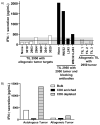
Figure 4. The treatment TIL exhibited autologous tumor recognition restricted by HLA class II that was enriched in CD4+ lymphocytes
IFNγ release was assayed by coculture and ELISA as described in Figure 2. Six allogeneic, HLA d mismatched FrTu were used as targets (left of figure) but failed to stimulate substantial IFNγ release. Three HLA-mismatched allogeneic TIL were cocultured with the 2950 FrTu but did not cause IFNγ release, demonstrating that the 2950 FrTu was not non-specifically stimulatory. The horizontal line at 200 pg/ml indicates background. The grey shaded area indicates one standard deviation of the non-specific values around the background. Autologous tumor was used with no antibody blocking, and with W6/32 (anti-Class I), L243 (anti-HLA-DR), or both W6/32 and L243. L243 significantly blocked tumor recognition by the autologous the autologous TIL. The specificity of antibody blocking was confirmed by inhibition of IFNγ release from TIL DMF5 (HLA- A2/MART1:27–35) and TIL E11 (HLA-DRB07/gp100:170–190) after co-culture with the melanoma cell line 624-CIITA.
Figure 5
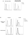
Figure 5. HLA class II was absent and was not inducible by IFNγ on the melanoma cell line derived from the recurrent pelvic tumor mass
A) HLA-DR was not expressed by the melanoma cell line derived from the recurrent pelvic mass while HLA class I (W6/32) was expressed (left). After forced expression by retroviral transduction with CIITA, HLA class II was observed while HLA class I was unchanged (right). B) The cell line 624 (upper) and the pelvic tumor line (lower) responded differently to IFNγ induction. HLA class I was expressed at all concentrations of IFNγ for both cell lines (left). HLA class II was induced by IFNγ in a dose dependent manner in the 624 cell line while no class II expression was induced at any IFNγ dose in the pelvic tumor line (right)

References

    1. Jemal A, Siegel R, Ward E, Hao Y, Xu J, Thun MJ. Cancer statistics, 2009. CA Cancer J Clin. 2009;59:225–49. - PubMed
    1. Rigel DS. Trends in dermatology: melanoma incidence. Arch Dermatol. 146:318. - PubMed
    1. Chapman PB, Hauschild A, Robert C, et al. Improved survival with vemurafenib in melanoma with BRAF V600E mutation. N Engl J Med. 364:2507–16. - PMC - PubMed
    1. Hodi FS, O’Day SJ, McDermott DF, et al. Improved survival with ipilimumab in patients with metastatic melanoma. N Engl J Med. 363:711–23. - PMC - PubMed
    1. Phan GQ, Yang JC, Sherry RM, et al. Cancer regression and autoimmunity induced by cytotoxic T lymphocyte-associated antigen 4 blockade in patients with metastatic melanoma. Proc Natl Acad Sci U S A. 2003;100:8372–7. - PMC - PubMed

MeSH terms