. 2012 Aug;40(15):7358-67.
doi: 10.1093/nar/gks407. Epub 2012 May 10.

The BLM helicase contributes to telomere maintenance through processing of late-replicating intermediate structures

Affiliations

The BLM helicase contributes to telomere maintenance through processing of late-replicating intermediate structures

Colleen Barefield et al. Nucleic Acids Res. 2012 Aug.

Abstract

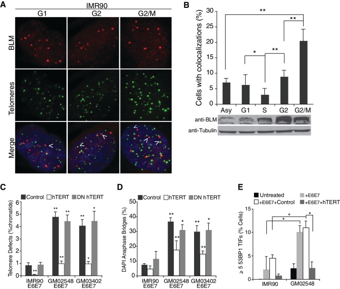
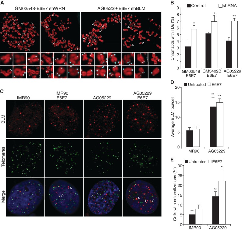
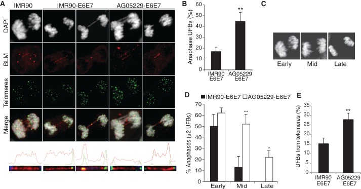
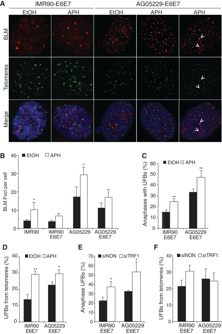
Werner's syndrome (WS) and Bloom's syndrome (BS) are cancer predisposition disorders caused by loss of function of the RecQ helicases WRN or BLM, respectively. BS and WS are characterized by replication defects, hyperrecombination events and chromosomal aberrations, which are hallmarks of cancer. Inefficient replication of the G-rich telomeric strand contributes to chromosome aberrations in WS cells, demonstrating a link between WRN, telomeres and genomic stability. Herein, we provide evidence that BLM also contributes to chromosome-end maintenance. Telomere defects (TDs) are observed in BLM-deficient cells at an elevated frequency, which is similar to cells lacking a functional WRN helicase. Loss of both helicases exacerbates TDs and chromosome aberrations, indicating that BLM and WRN function independently in telomere maintenance. BLM localization, particularly its recruitment to telomeres, changes in response to replication dysfunction, such as in WRN-deficient cells or after aphidicolin treatment. Exposure to replication challenge causes an increase in decatenated deoxyribonucleic acid (DNA) structures and late-replicating intermediates (LRIs), which are visible as BLM-covered ultra-fine bridges (UFBs) in anaphase. A subset of UFBs originates from telomeric DNA and their frequency correlates with telomere replication defects. We propose that the BLM complex contributes to telomere maintenance through its activity in resolving LRIs.


Figures

Figure 1.
Figure 1.
BLM localizes to a subset of telomeres preferentially in G2/M. (A) IF-FISH images of synchronized IMR90 cells, using a BLM specific antibody (red) and a telomeric probe, FITC-[CCCTAA]4 (green). Arrows indicate colocalization events between BLM and telomeric foci. (B) Top panel: quantification of IF-FISH data, showing the percentage of IMR90 cells with over two colocalization events. At least 100 cells were counted per time-point and values were averaged from four independent experiments. Error bars represent the standard deviation and P-values were calculated with a Student’s t-test (*P < 0.05 and **P < 0.005). Bottom panel: western blot of synchronized IMR90 cells probed with BLM antibody or a tubulin control. IMR90 lysates were collected at the indicated stages of the cell cycle. (C) Percentage of chromatids with TDs in IMR90 or BS fibroblast cell lines expressing an empty vector control, hTERT or DN_hTERT. At least 1500 chromatids per cell line, per experiment were counted from three independent experiments. (D) DAPI-positive anaphase bridges in IMR90 and BS fibroblast cells expressing an empty vector control, hTERT or DN_hTERT. At least 55 anaphases per cell line were counted from three independent experiments. (E) Percentage of IMR90 and GM02548 BS cells with over five TIF. Cells were fixed with 4% PFA and prepared for IF-FISH with a 53BP1-specific antibody and the telomeric probe FITC-[CCCTAA]4. At least 100 cells from each cell line were counted in two independent experiments. Error bars represent the standard deviation and P-values were calculated with a Student’s t-test (*P < 0.05 and **P < 0.005).
Figure 2.
Figure 2.
Loss of both BLM and WRN RecQ helicases exacerbates TDs. (A) Images of FISH of metaphase chromosomes from GM02548-E6E7 (BS) fibroblasts after WRN knockdown and AG05229-E6E7 (WS) fibroblasts after BLM knockdown. Arrows indicate chromosomes in the magnified panels below each metaphase spread. (B) Percentage of chromatids with TDs. At least 2000 chromatids from each cell line were counted, and average values were compiled from two independent experiments. (C) IMR90 and WS cells were fixed with 4% PFA and used for IF-FISH with a BLM antibody and the telomeric probe FITC-[CCCTAA]4. IF-FISH images of indicated fibroblast cell lines, showing BLM (red) and telomeres (green); arrows show colocalization events. (D) Average number of BLM foci per cell and (E) percentage of cells with more than two colocalization events between BLM and telomeres. The values for each cell line were averaged from three independent IF-FISH experiments. Arrow bars represent standard deviation, and P-values were calculated by a Student’s t-test (*P < 0.05 and **P < 0.005).
Figure 3.
Figure 3.
Ultra-fine bridges (UFBs) are more frequent and persist longer in WS fibroblasts. (A) IF-FISH images of UFB-positive anaphases representing UFBs that do not localize with telomeric foci (left panel), UFBs that colocalize with a telomeric foci at one end or at the center (second and third panels) and UFBs that colocalize with telomeres at both ends (final two panels). Graphs illustrating the signal intensities generated by the profile function of the AxioVision software are shown below each anaphase. (B) The percentage of anaphases with at least one UFB and (C) DAPI-stained anaphases representing three stages of anaphase, determined by distance (in µm) between dividing cells: early, mid and late anaphases. (D) Percentage of UFB-positive anaphases with multiple UFBs per anaphase. Data from IMR90-E6E7 and AG05229-E6E7 cells were grouped according to progress through anaphase (panel C). At least 25 anaphases were analyzed for each cell line per experiment, from four independent experiments. Arrow bars show standard deviation, and P-values were calculated by a Student’s t-test (*P < 0.05 and **P < 0.005). (E) The percentage of UFBs extending from telomeres (T-UFBs). The values were calculated from four independent IF-FISH experiments, where at least 30 anaphases were analyzed from each cell line.
Figure 4.
Figure 4.
Replication challenge increases UFB formation. (A) IF-FISH images from IMR90-E6E7 and AG05229-E6E7 fibroblast cells. Arrows indicate colocalization events at BLM bridges observed in interphase cells. (B) Average number of BLM foci per cell. At least 100 cells were counted per condition from three independent experiments. (C) Percentage UFB-positive anaphases and (D) percentage of UFBs extending from telomeres (T-UFBs) in IMR90-E6E7 and AG05229-E6E7 cells treated with ethanol or aphidicolin. At least 30 anaphases were counted per condition from three independent experiments. (E) Percentage of UFBs and (F) UFBs that extend from telomeric foci (T-UFBs) on TRF1 suppression. At least 35 anaphases were counted per cell line from two independent experiments. Arrow bars show standard deviation, and P-values were calculated from a Student’s t-test (*P < 0.05 and **P < 0.005).

References

    1. de Lange T. Protection of mammalian telomeres. Oncogene. 2002;21:532–540. - PubMed
    1. de Lange T. Shelterin: the protein complex that shapes and safeguards human telomeres. Genes Dev. 2005;19:2100–2110. - PubMed
    1. O'Sullivan RJ, Karlseder J. Telomeres: protecting chromosomes against genome instability. Nat. Rev. Mol. Cell Biol. 2010;11:171–181. - PMC - PubMed
    1. Palm W, de Lange T. How shelterin protects mammalian telomeres. Annu. Rev. Genet. 2008;42:301–334. - PubMed
    1. Opresko PL, Cheng WH, von Kobbe C, Harrigan JA, Bohr VA. Werner syndrome and the function of the Werner protein; what they can teach us about the molecular aging process. Carcinogenesis. 2003;24:791–802. - PubMed

Publication types

MeSH terms