. 2012 Jun 29;287(27):22497-508.
doi: 10.1074/jbc.M111.330589. Epub 2012 May 10.

Neural transmembrane protease and endothelial Gs protein activation in cell contact-dependent signaling between neural stem/progenitor cells and brain endothelial cells

Affiliations

Neural transmembrane protease and endothelial Gs protein activation in cell contact-dependent signaling between neural stem/progenitor cells and brain endothelial cells

Hsiu-Hui Tung et al. J Biol Chem. .

Abstract

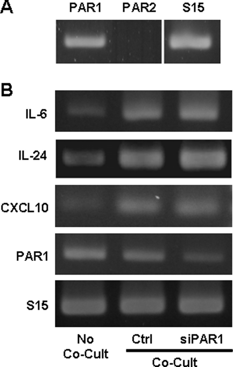
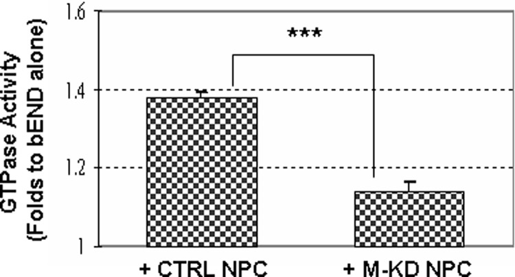
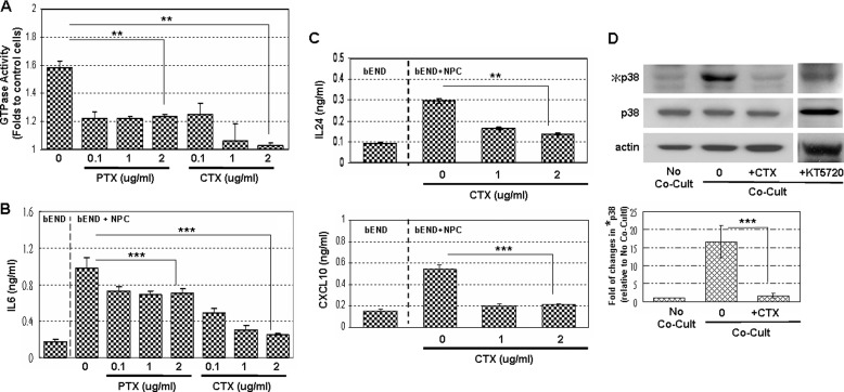
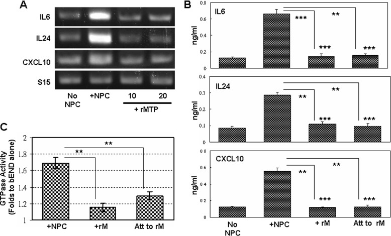
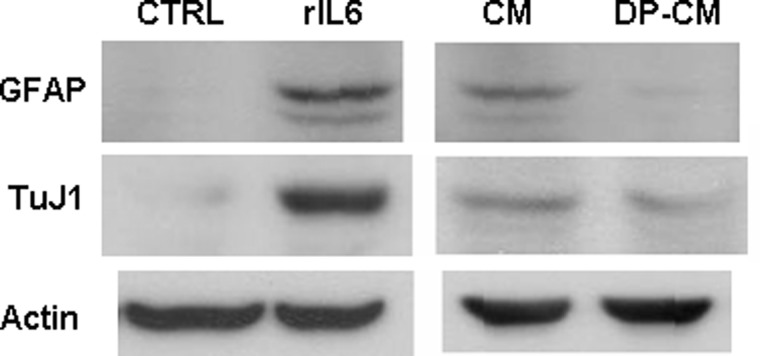
Vasculature is an important component of the neural stem cell niche in brain. It regulates neural stem/progenitor (NS/P) cell self-renewal, differentiation, and migration. In the neurogenic niches of adult brain, NS/P cells lie close to blood vessels, and proliferating NS/P cells frequently contact the vasculature. In the present study we showed that NS/P cells in co-culture with brain endothelial (bEND) cells activated endothelial G proteins and p38 mitogen-activated protein kinase (p38 MAPK) and stimulated cytokine/chemokine expression. These NS/P cell-induced endothelial responses took place during NS/P cell and bEND cell direct contact and were critically dependent on the expression of the type II transmembrane serine protease matriptase (MTP) by NS/P cells, because knocking down of MTP in NS/P cells impaired and re-expression of MTP restored their ability to induce endothelial cytokine/chemokine expression, p38 MAPK, or G protein activation. Cholera toxin blocked NS/P cell-induced endothelial responses, suggesting that the endothelial G protein activated by NS/P MTP is in the G(s) subfamily. The addition of p38 MAPK inhibitor impaired NS/P cell-induced endothelial cytokine/chemokine expression. The known G protein-coupled receptor substrate of MTP, protease-activated receptor 2, was not involved in this system. These results revealed a novel signaling pathway in neural stem cell vascular niches that is mediated by neural MTP and endothelial G(s) protein signaling at the cell-cell interface. This is the first report of direct cell-cell signaling between NS/P and bEND cells.


Figures

FIGURE 1.
FIGURE 1.
NS/P cells induced cytokine/chemokine expression in bEND cells through cell contact. A, RT-PCR assay of IL6, IL24, and CXCL10 mRNA in NS/P cells (NPC) or bEND cells (bEND) cultured either separately (No Co-Cult) or co-cultured under direct contact conditions (Co-Cult). NS/P marker nestin (Nestin) and endothelial marker FLK1 indicate sufficient purity of bEND cells isolated from co-culture. B, RT-PCR assay of IL6, IL24, and CXCL10 mRNA. C, ELISA of IL6, IL24, and CXCL10 proteins released by bEND cells cultured alone (No Co-Cult) or co-cultured (Co-Cult) with NS/P cells under direct contact conditions (Cont) or in a Transwell (N-Cont). The asterisks above the bars indicate a significant difference of each co-cultured condition compared with No Co-Cult. Comparison was also made between two co-cultured conditions. ***, p ≦ 0.001. D, RT-PCR of IL6, IL24, and CXCL10 in bEND cells, in NS/P cells isolated from the SVZ of adult mouse brain lateral ventricle (SVZ) and in NS/P cells of neural cortex from 14-day mouse embryo (E14) after contact co-culture (Co-Cult). Expression of IL6, IL24, and CXCL10 in bEND cells cultured without NS/P cells is also shown. Mouse small ribosome protein 15 (S15) in each figure serves as an internal loading control for RT-PCR. The same results for RT-PCR were observed in at least three independent experiments.
FIGURE 2.
FIGURE 2.
NS/P cells induce endothelial p38 MAPK activation for cytokine/chemokine production. A, cell lysates of bEND cells (bEND) and NS/P cells (NPC) that were either cultured separately (No) or under contact conditions (Co-Cult) were assayed by Western blot for the expression of the total p38 MAPK (p38), phosphorylated p38 MAPK (*p38), total PKCα (PKC) or phosphorylated PKCα (*PKC). Actin is the endogenous loading control. B, immunostaining of phosphorylated p38 MAPK (p38*, red fluorescence) in NS/P cells-bEND cells contact co-culture. Anti-GFP was used to show NS/P cells (GFP, green fluorescence). Cell nuclei were stained with DAPI (blue fluorescence). Arrowheads indicate bEND cells that are in contact with NS/P cells; arrows indicate bEND cells that are not in contact with NS/P cells. The two images in the box on the bottom left are bEND cells cultured either without NS/P cells (left panel) or co-cultured with NS/P cells in a Transwell (right panel). Arrows in these images indicate basal *p38 staining in bEND cells. The cells stained with control immunoglobins of the same subtype were also shown (control Ig). The scale bars are 50 μm. C, RT-PCR of IL6, IL24, or CXCL10 in bEND cells cultured without NS/P cells (No Co-Cult), in contact with NS/P cells (Co-Cult, Ctrl), or in contact with NS/P cells in the presence of SB203580 (+p38I). S15 is an internal loading control. The graph shows quantitation of RT-PCR by densitometry. The values of each molecule in co-culture were normalized to that of No Co-Cult that is set at 1. Comparisons were made between control and SB203580 (+p38I) co-culture. *, p ≦ 0.05.
FIGURE 3.
FIGURE 3.
Endogenous expression of MTP in NS/P cells mediates cell-contact induced endothelial p38 MAPK activation and cytokine/chemokine expression. A, RT-PCR assay of MTP and IL6 mRNAs in NS/P cells (NPC), bEND cells (bEND), and MTP knockdown NS/P cells (M-KD NPC) without co-culture (No Co-Cult). MTP and IL6 mRNAs in bEND cells in contact with NS/P cells transfected with control siRNA (CT Co-Cult) or with MTP siRNA (KD Co-Cult) were compared. Endothelial FLK1 and NPC marker nestin are used as monitors for bEND cells and their purity after co-culture. B, expression of CXCL10 or IL24 in bEND cells cultured without NS/P cells (No Co-Cult), after direct contact co-culture (Co-Cult) with control siRNA transfected (Ctrl) or with MTP siRNA transfected (M-KD) NS/P cells. C, semiquantitative PCR for IL6, IL24, and CXCL10 in bEND cells cultured without NS/P cells (No Co-Cult), after direct contact co-culture with control siRNA transfected NS/P cells (Ctrl Co-Cult) or with MTP siRNA transfected NS/P cells (M-KD Co-Cult). PCR product after 25, 26, and 27 cycles of amplification were shown. The PCR product of internal control S15 was taken after 20, 21, and 22 cycles of amplification. A–C show the representative data of at least three independent experiments. S15 is loading control for RT-PCR. D, ELISA of IL6, IL24, and CXCL10 proteins in conditioned medium of bEND cells cultured without NS/P cells (No NPC), bEND cells after contact with control siRNA transfected (+Ctrl NPC), or with MTP knockdown (+M-KD NPC) NS/P cells. Asterisks above the bars indicate significant difference compared with No NPC. Comparisons were also made between +CTRL NPC and +M-KD NPC. E, total and phosphorylated p38 MAPK protein (p38 and *p38, respectively) in bEND cells cultured in conditions described in D. Actin is the endogenous loading control for Western blot. The graph shows quantitation of chemiluminescence by densitometry. The value of phosphorylated p38 was normalized to that of the total p38 for each sample; the resulting value was then normalized to that of bEND cells without co-culture that is set at 1. Comparisons were made between +CTRL NPC and +M-KD NPC. F, image on the left shows RT-PCR of MTP in NS/P cells (Ctrl), in MTP knockdown NS/P cells (M-KD), and in MTP knockdown or control NS/P cells transfected with MTP expression vector (M-KD/pM and pM, respectively). The graphs on the right show ELISA of IL6, IL24, and CXCL10 proteins in conditioned medium of bEND cells cultured with MTP knockdown (M-KD) cells and with MTP knockdown or control NS/P cells carrying MTP expression vector (M-KD/pM and pM, respectively). The dark dotted line across each graph shows the amount of each molecule induced by the control NS/P cells. Cultures with M-KD/pM or pM were compared with cultures with M-KD. **, p ≦ 0.01.
FIGURE 4.
FIGURE 4.
Ecotopic expression of MTP in bEND cells induces their p38 MAPK activation and cytokine/chemokine expression. A, Western immunoblot assay of MTP protein, phosphorylated (*p38), and total (p38) p38 MAPK protein in control bEND cells transfected with an empty vector (Ctrl) or bEND cells transfected with MTP-expressing plasmid (pMTP). The graph shows quantitation of chemiluminescence by densitometry. The value of phosphorylated p38 was normalized to that of the total p38 for each sample; the resulting value was then normalized to that of control bEND cells (set at 1). B, RT-PCR of MTP, IL6, IL24, and CXCL10 mRNA in bEND cells collected in A. S15 and actin are loading controls for RT-PCR and Western blot, respectively. The same results were observed in at least three independent experiments. C, ELISA of the secreted IL6 protein in bEND cells collected in A. **, p ≦ 0.01.
FIGURE 5.
FIGURE 5.
PAR1 or PAR2 is not involved in NS/P-bEND cell contact induced cytokine/chemokine production. A, RT-PCR of PAR1 and PAR2 in bEND cells. B, RT-PCR of IL6, IL24, and CXCL10 mRNA in bEND cells cultured without NS/P cells (No Co-Cult), in bEND cells (Ctrl), or PAR1 knockdown bEND cells (siPAR1) after contact with NS/P cells (Co-Cult). RT-PCR of PAR1 shows the knockdown efficiency. S15 is an internal loading control. Shown here are representative data of three independent experiments.
FIGURE 6.
FIGURE 6.
NS/P cell-induced endothelial GTPase activity in contact co-culture is dependent on endogenous expression of MTP in NS/P cells. GTPase activity in bEND cells after contact with control siRNA transfected (CTRL) or with MTP siRNA transfected NS/P cells (M-KD) were normalized to that in bEND cells cultured without NS/P cells (set at 1). ***, p ≦ 0.001.
FIGURE 7.
FIGURE 7.
CTX-sensitive endothelial G protein is responsible for NS/P cell-induced endothelial IL6, IL24, and CXCL10 production and p38 MAPK activation. A, endothelial GTPase activity in bEND cells co-cultured with NS/P cells in the absence (0) or presence of various concentrations (0.1, 1, and 2 μg/ml) of PTX or CTX. The data were normalized to bEND cells cultured without NS/P cells (set at 1). B and C, ELISA of IL6 protein (B) and ELISA of IL24 and CXCL10 in bEND cells (C) cultured without NS/P cells (bEND) or in contact with NS/P cells (bEND+NPC) in the absence (0) or presence of various concentrations (0.1, 1, and 2 μg/ml) of PTX or CTX. D, Western immunoblot of p38 MAPK in bEND cells cultured alone (No Co-Cult) or with NS/P cells (Co-Cult) in the absence (0) or presence of 1 ng/ml CTX (+CTX) or KT5720 (+KT5720). Actin is the internal loading control for Western blot assay. The graph shows quantitation of chemiluminescence by densitometry. The value of phosphorylated p38 was normalized to that of the total p38 for each sample; the resulting value was then normalized to that of bEND cells without co-culture that is set at 1. Toxin-treated cultures were compared with zero-toxin-treated cultures. **, p ≦ 0.01; ***, p ≦ 0.001.
FIGURE 8.
FIGURE 8.
Recombinant MTP protease fails to induce brain endothelial responses. A, RT-PCR of IL6, IL24, and CXCL10 mRNA in bEND cells cultured without NS/P cells (No Co-Cult), in contact with NS/P cells (Co-Cult), or in medium containing recombinant protein of MTP protease domain (+rMTP, 10 or 20 nm). Shown here are representative data of three independent experiments. B, ELISA of IL6, IL24, and CXCL10 proteins in conditioned medium of bEND cells cultured without NS/P cells (No NPC), after contact with NS/P cells (+NPC), cultured in medium containing recombinant protein of MTP protease domain (+rM), or cultured on dishes coated with recombinant protein of MTP protease domain (Att to rM). Recombinant protein-treated cultures (+rM and Att to rM) were compared with No NPC cultures (asterisks above bars) and to +NPC cultures. C, GTPase activity in bEND cells after contact with NS/P cells (+NPC), cultured in medium containing recombinant protein of MTP protease domain (+rM), or cultured on dishes coated with recombinant protein of MTP protease domain (Att to rM). The data were normalized to bEND cells cultured without NPC (set at 1). The cells cultured with recombinant protein (+rM or Att to rM) were compared with cells cultured with NS/P cells (+NPC). **, p ≦ 0.01; ***, p ≦ 0.001.
FIGURE 9.
FIGURE 9.
NS/P cell-induced endothelial IL6 promotes glial and neural differentiation of NS/P cells. Expression of glial protein GFAP and early neuron protein TuJ1 in NS/P cells cultured under differentiation conditions in the absence (CTRL) or presence of recombinant IL6 protein (rIL6) or cultured with conditioned medium collected of NS/P-bEND cells in contact co-culture before (CM) or after (DP-CM) removal of IL6 protein by specific antibody. Actin is the internal loading control for Western blot assay.

References

    1. Temple S. (2001) The development of neural stem cells. Nature 414, 112–117 - PubMed
    1. Zerlin M., Goldman J. E. (1997) Interactions between glial progenitors and blood vessels during early postnatal corticogenesis. Blood vessel contact represents an early stage of astrocyte differentiation. J. Comp. Neurol. 387, 537–546 - PubMed
    1. Alvarez-Buylla A., Lim D. A. (2004) For the long run. Maintaining germinal niches in the adult brain. Neuron 41, 683–686 - PubMed
    1. Palmer T. D., Willhoite A. R., Gage F. H. (2000) Vascular niche for adult hippocampal neurogenesis. J. Comp. Neurol. 425, 479–494 - PubMed
    1. Tavazoie M., Van der Veken L., Silva-Vargas V., Louissaint M., Colonna L., Zaidi B., Garcia-Verdugo J. M., Doetsch F. (2008) A specialized vascular niche for adult neural stem cells. Cell Stem Cell 3, 279–288 - PMC - PubMed

Publication types

MeSH terms