

Outcome of fetuses with diagnosis of isolated short femur in the second half of pregnancy
- PMID: 22577572
- PMCID: PMC3345228
- DOI: 10.5402/2012/268218
Outcome of fetuses with diagnosis of isolated short femur in the second half of pregnancy
Abstract
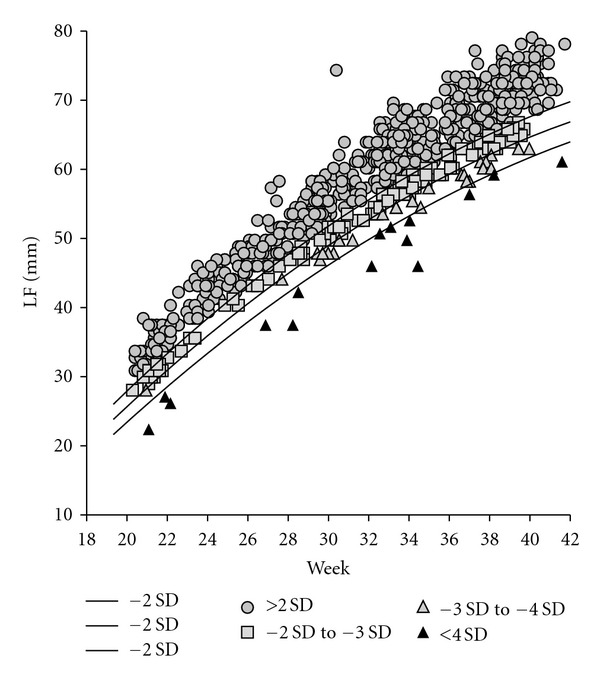
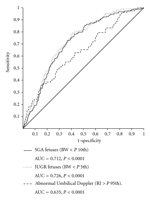
Objectives. To assess the outcome of fetuses with isolated short femur detected at 19-41 weeks and determine to what extent this incidental finding should be a cause of concern in fetuses with a normal previous follow-up. Methods. 156 fetuses with isolated short femur were compared with a control group of 637 fetuses with normal femur length. FL values were converted into Z-scores and classified into 4 groups: control group: Z-score over -2, group 1: Z-score between -2 and -3, group 2: Z-score between -3 and -4, and group 3: Z-score below -4. FL values were plotted with the curves representing Z-scores -2, -3, and -4. To assess fetal outcome, the frequency of SGA, IUGR, abnormal umbilical Doppler (AUD), Down's syndrome, and skeletal dysplasia was determined for each group after delivery, and the relative risk in comparison with the control group was obtained. Finally, ROC curves were drawn in order to evaluate the FL diagnostic ability for the conditions appearing with increased frequency. Results. SGA, IUGR, and AUD were more frequent in the fetuses with short femur. Conversely, none of them presented Down's syndrome or skeletal dysplasia. According to ROC analysis, FL measurement behaved as a good diagnostic test for SGA and IUGR. Conclusions. A short femur diagnosis in a fetus with an otherwise normal follow-up determines just a higher risk of being small (SGA or IUGR).
Figures
References
-
- Carrascosa Lezcano A, Ferrández Longás A, Yeste Fernández D, et al. Estudio transversal español de crecimiento 2008. Parte I: valores de peso y longitud en recién nacidos de 26–42 semanas de edad gestacional. Anales de Pediatria. 2008;68(6):544–551. - PubMed
-
- Kurmanavicius J, Florio I, Wisser J, et al. Reference resistance indices of the umbilical, fetal middle cerebral and uterine arteries at 24–42 weeks of gestation. Ultrasound in Obstetrics and Gynecology. 1997;10(2):112–120. - PubMed
-
- Nyberg DA, Resta RG, Hickok DE, Hollenbach KA, Luthy DA, Mahony BS. Femur length shortening in the detection of Down syndrome: is prenatal screening feasible? American Journal of Obstetrics and Gynecology. 1990;162(5):1247–1252. - PubMed
-
- Kagan KO, Wright D, Baker A, Sahota D, Nicolaides KH. Screening for trisomy 21 by maternal age, fetal nuchal translucency thickness, free beta-human chorionic gonadotropin and pregnancy-associated plasma protein-A. Ultrasound in Obstetrics and Gynecology. 2008;31(6):618–624. - PubMed
-
- Goncalves L, Jeanty P. Fetal biometry of skeletal dysplasias: a multicentric study. Journal of Ultrasound in Medicine. 1994;13(12):977–985. - PubMed
LinkOut - more resources
Full Text Sources
