

Expression dynamics of Toll-like receptors mRNA and cytokines in porcine peripheral blood mononuclear cells stimulated by bacterial lipopolysaccharide
- PMID: 22578850
- PMCID: PMC11141511
- DOI: 10.1016/j.vetimm.2012.04.020
Expression dynamics of Toll-like receptors mRNA and cytokines in porcine peripheral blood mononuclear cells stimulated by bacterial lipopolysaccharide
Abstract
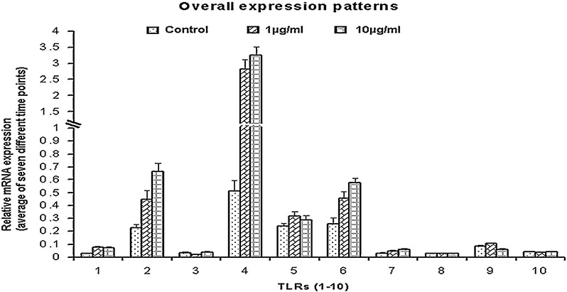
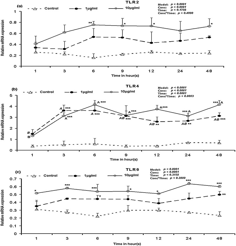
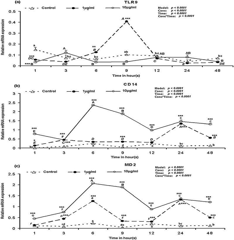
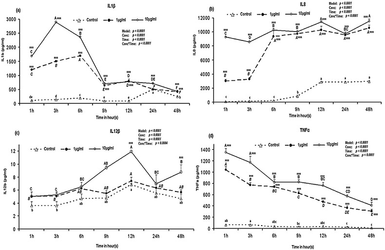
The Toll-like receptor (TLR)4 is critical for the recognition of Gram-negative bacterial lipopolysaccharide (LPS) but in porcine peripheral blood mononuclear cells (PBMCs) it may cooperate with other TLRs and lead to the production of inflammatory cytokines. Therefore, we analyzed TLR1-10 mRNA expression in porcine PBMCs stimulated with LPS over time (1-48 h) by using quantitative real-time PCR and cytokine proteins level by ELISA in culture supernatant. TLR1-10 mRNA was detectable in porcine PBMCs. When compared with the control (non-stimulated), TLR1 mRNA were increased (p<0.05) at 3 h after challenge with 1 μg/ml LPS, whereas TLR1 and TLR2 mRNA were increased (p<0.01) at 6 h after challenge with 10 μg/ml LPS. TLR4 increased (p<0.001) at 3h after challenge with LPS and remained constant. TLR5 and TLR6 mRNA increased (p<0.05) at 9 h and 1 h after of LPS stimulation, respectively. The mRNA of CD14 and MD2 were increased (p<0.001) at 1h after LPS stimulation. Additionally, at most of the time analyzed, the mRNA expression increased with the dose of LPS. The LPS concentration had influence (p<0.05) on all the TLRs expression except TLR10; whereas time had effect (p<0.05) on all TLRs expression except TLR2, 3, 6 and 10. When compared to the control, the cytokines IL1b, IL8 and TNFα proteins were increased (p<0.001) immediately at 1 h after LPS stimulation and remained constant till 48 h. IL12b was increased (p<0.001) 12 h after challenge with 10 μg/ml of LPS. Although IL8 level was the highest, the higher (p<0.05) expression of all these inflammatory cytokines indicate that upon interacting with TLRs, LPS exerted inflammatory response in PBMCs through the production of Th1 type cytokines. The production of cytokines was influenced (p<0.001) by both the dose of LPS and the stimulation time. Hence, the porcine PBMCs are likely able to express all members of TLRs.
Copyright © 2012 Elsevier B.V. All rights reserved.
Figures
Similar articles
-
Expression and function of toll-like receptors in human circulating endothelial colony forming cells.Immunol Lett. 2015 Nov;168(1):98-104. doi: 10.1016/j.imlet.2015.09.014. Epub 2015 Oct 8. Immunol Lett. 2015. PMID: 26433057
-
Increased responsiveness to toll-like receptor 4 stimulation in peripheral blood mononuclear cells from patients with recent onset rheumatoid arthritis.Mediators Inflamm. 2008;2008:132732. doi: 10.1155/2008/132732. Mediators Inflamm. 2008. PMID: 18584044 Free PMC article.
-
Differential responses between monocytes and monocyte-derived macrophages for lipopolysaccharide stimulation of calves.Cell Mol Immunol. 2009 Jun;6(3):223-9. doi: 10.1038/cmi.2009.30. Cell Mol Immunol. 2009. PMID: 19567206 Free PMC article.
-
Expression of Toll-like receptors and downstream genes in lipopolysaccharide-induced porcine alveolar macrophages.Vet Immunol Immunopathol. 2012 Mar 15;146(1):62-73. doi: 10.1016/j.vetimm.2012.02.001. Epub 2012 Feb 6. Vet Immunol Immunopathol. 2012. PMID: 22365308
-
Possible involvement of toll-like receptor 4 in endothelial cell activation of larger vessels in response to lipopolysaccharide.Pathobiology. 2002;70(1):18-25. doi: 10.1159/000066000. Pathobiology. 2002. PMID: 12415188
Cited by
-
Kinetics of the West Nile virus induced transcripts of selected cytokines and Toll-like receptors in equine peripheral blood mononuclear cells.Vet Res. 2016 Jun 7;47(1):61. doi: 10.1186/s13567-016-0347-8. Vet Res. 2016. PMID: 27267361 Free PMC article.
-
A comparative review of toll-like receptor 4 expression and functionality in different animal species.Front Immunol. 2014 Jul 10;5:316. doi: 10.3389/fimmu.2014.00316. eCollection 2014. Front Immunol. 2014. PMID: 25071777 Free PMC article. Review.
-
Interaction between Leptospiral Lipopolysaccharide and Toll-like Receptor 2 in Pig Fibroblast Cell Line, and Inhibitory Effect of Antibody against Leptospiral Lipopolysaccharide on Interaction.Asian-Australas J Anim Sci. 2015 Feb;28(2):273-9. doi: 10.5713/ajas.14.0440. Asian-Australas J Anim Sci. 2015. PMID: 25557825 Free PMC article.
-
Porcine FcεRI Mediates Porcine Reproductive and Respiratory Syndrome Virus Multiplication and Regulates the Inflammatory Reaction.Virol Sin. 2018 Jun;33(3):249-260. doi: 10.1007/s12250-018-0032-3. Epub 2018 May 14. Virol Sin. 2018. PMID: 29761267 Free PMC article.
-
The porcine innate immune system: an update.Dev Comp Immunol. 2014 Aug;45(2):321-43. doi: 10.1016/j.dci.2014.03.022. Epub 2014 Apr 4. Dev Comp Immunol. 2014. PMID: 24709051 Free PMC article. Review.
References
-
- Akashi-Takamura S, Miyake K, 2008. TLR accessory molecules. Curr. Opin. Immunol 20, 420–425. - PubMed
-
- Akira S, Takeda K, 2004. Toll-like receptor signalling. Nat. Rev. Immunol 4, 499–511. - PubMed
-
- Alvarez B, Revilla C, Domenech N, Perez C, Martinez P, Alonso F, Ezquerra A, Domiguez J, 2008. Expression of toll-like receptor 2 (TLR2) in porcine leukocyte subsets and tissues. Vet. Res 39. - PubMed
-
- Burkey TE, Skjolaas KA, Dritz SS, Minton JE, 2009. Expression of porcine Toll-like receptor 2, 4 and 9 gene transcripts in the presence of lipopolysaccharide and Salmonella enterica serovars Typhimurium and Choleraesuis. Vet. Immunol. Immunopathol 130, 96–101. - PubMed
MeSH terms
Substances
Grants and funding
LinkOut - more resources
Full Text Sources
Research Materials
