. 2012 May 11;149(4):753-67.
doi: 10.1016/j.cell.2012.04.017.

Cell-free formation of RNA granules: low complexity sequence domains form dynamic fibers within hydrogels

Affiliations

Cell-free formation of RNA granules: low complexity sequence domains form dynamic fibers within hydrogels

Masato Kato et al. Cell. .

Abstract

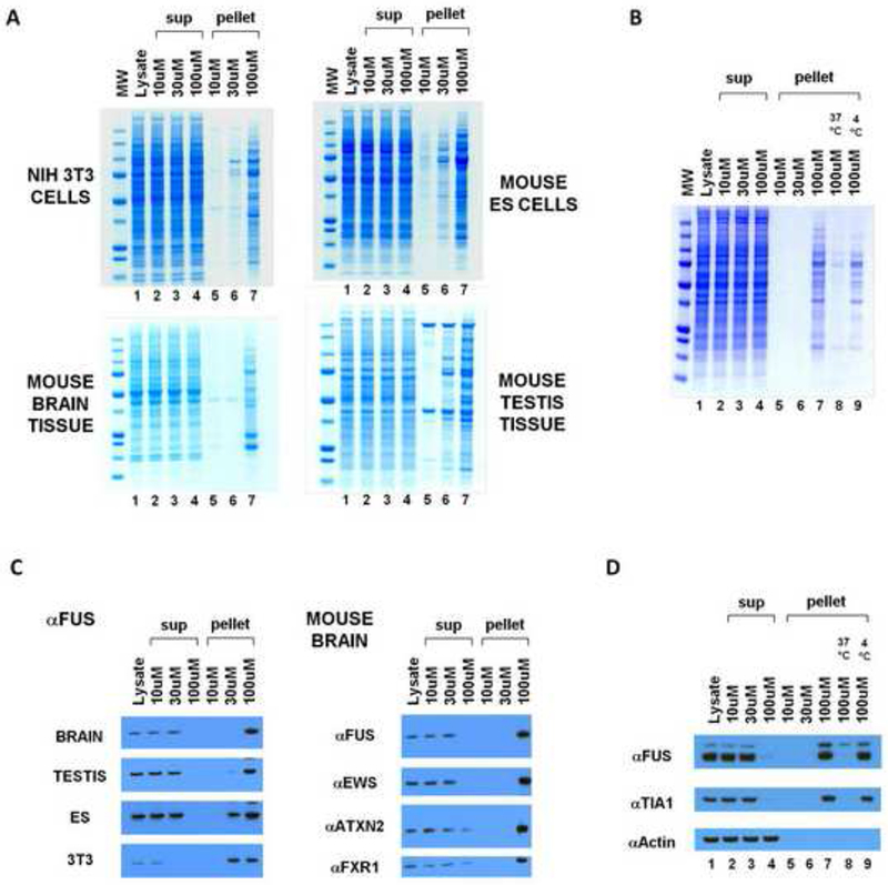
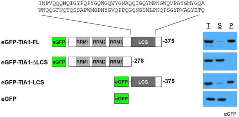
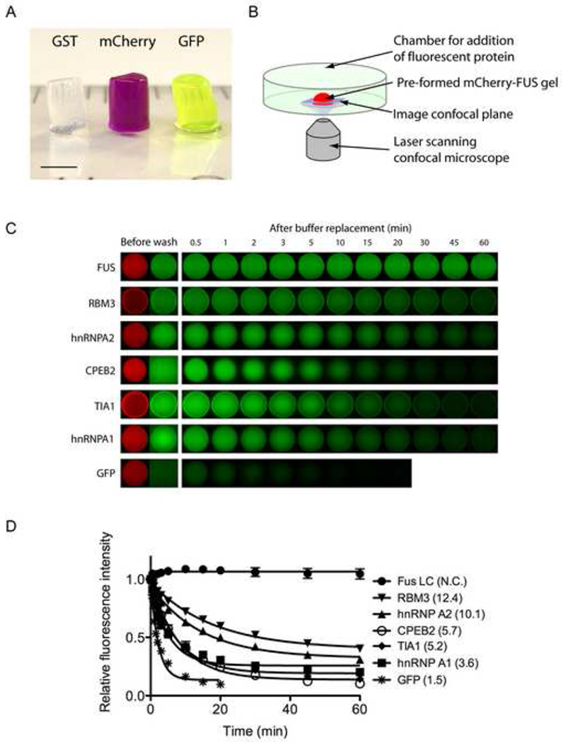
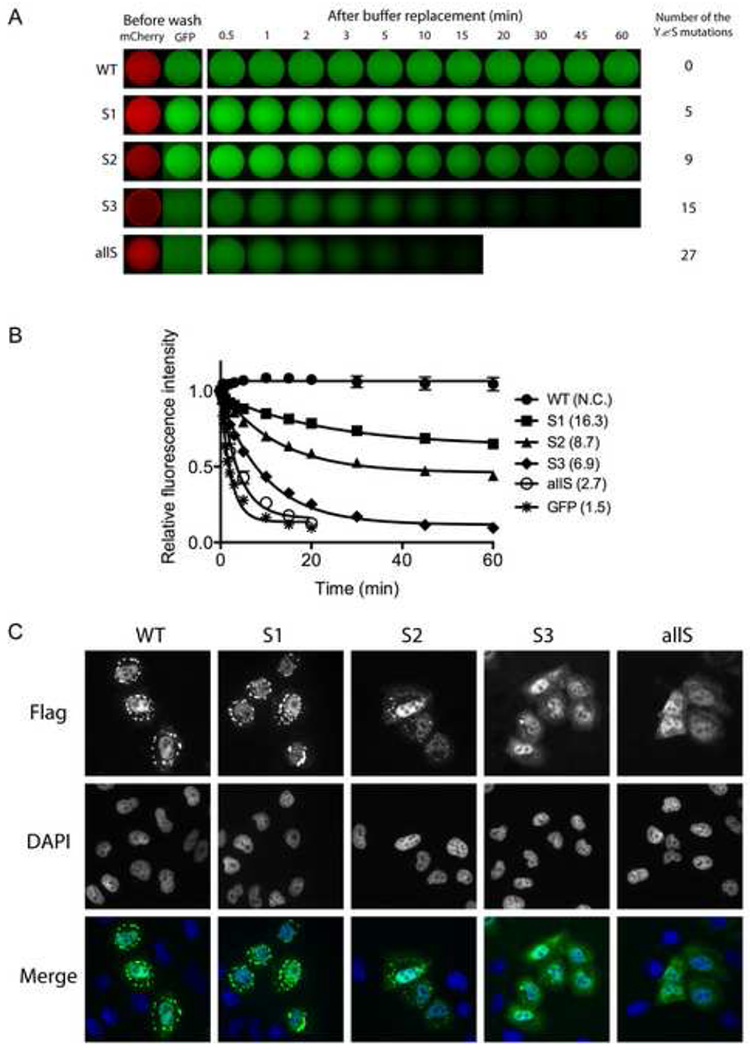
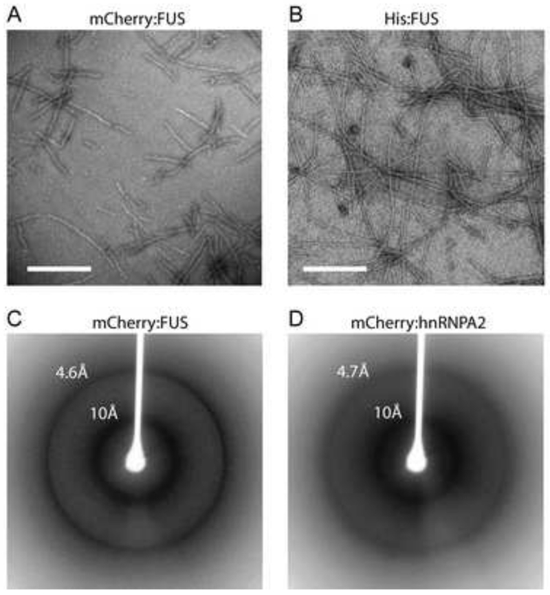
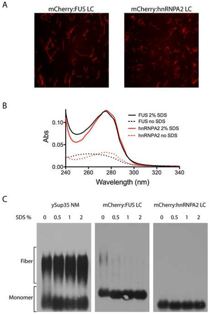
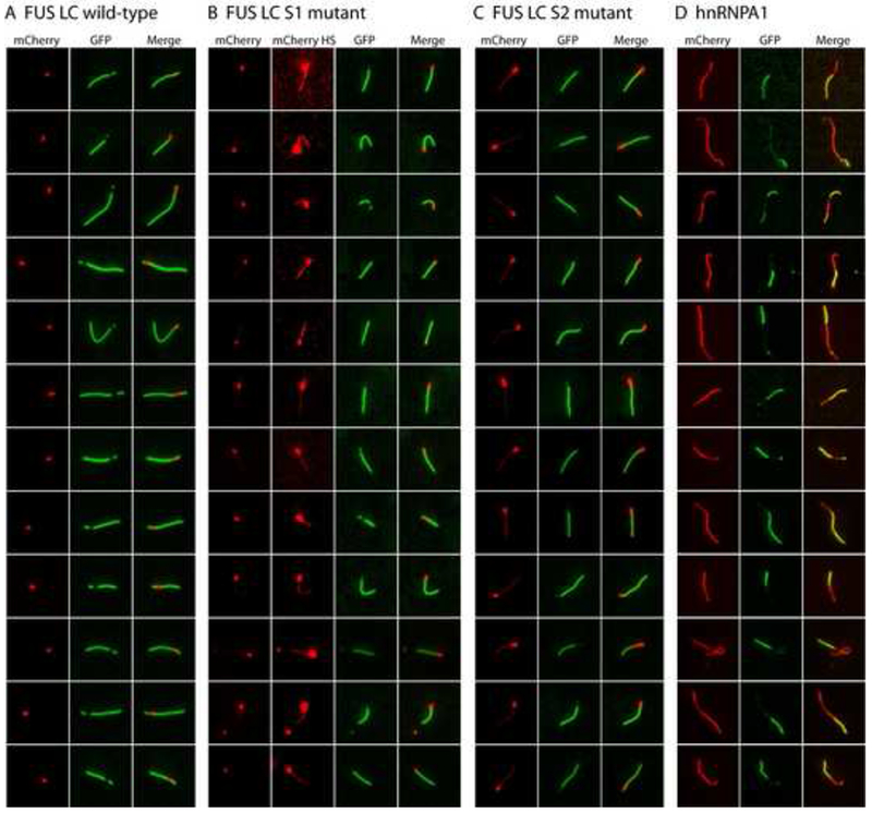
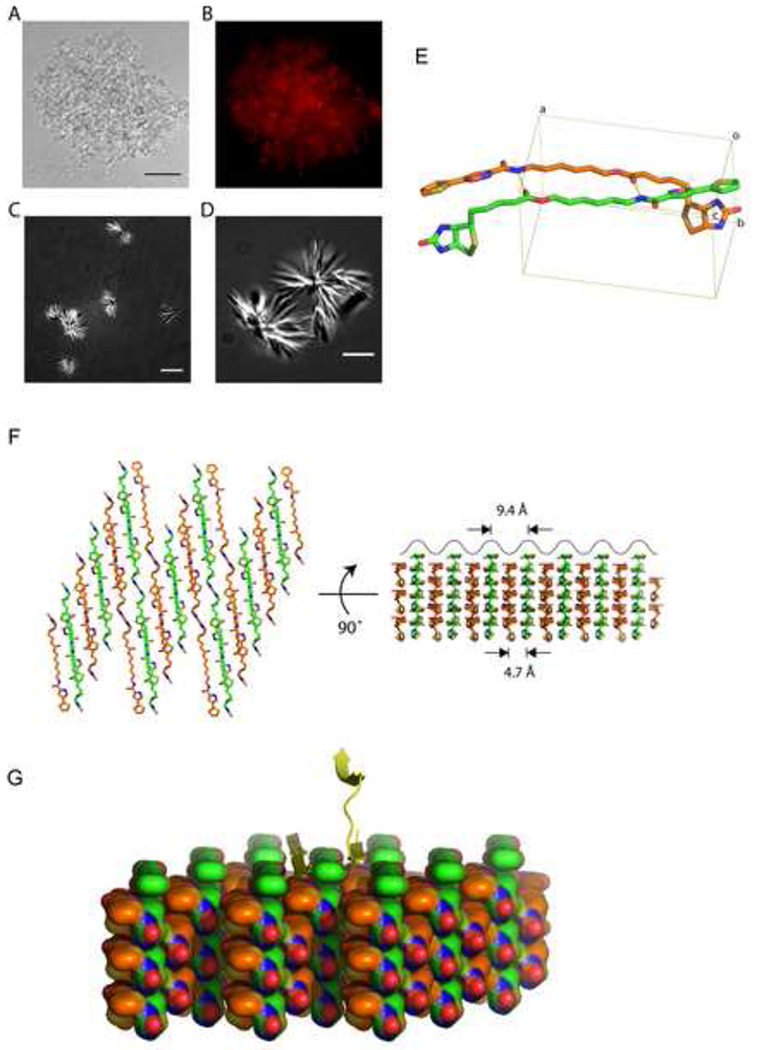
Eukaryotic cells contain assemblies of RNAs and proteins termed RNA granules. Many proteins within these bodies contain KH or RRM RNA-binding domains as well as low complexity (LC) sequences of unknown function. We discovered that exposure of cell or tissue lysates to a biotinylated isoxazole (b-isox) chemical precipitated hundreds of RNA-binding proteins with significant overlap to the constituents of RNA granules. The LC sequences within these proteins are both necessary and sufficient for b-isox-mediated aggregation, and these domains can undergo a concentration-dependent phase transition to a hydrogel-like state in the absence of the chemical. X-ray diffraction and EM studies revealed the hydrogels to be composed of uniformly polymerized amyloid-like fibers. Unlike pathogenic fibers, the LC sequence-based polymers described here are dynamic and accommodate heterotypic polymerization. These observations offer a framework for understanding the function of LC sequences as well as an organizing principle for cellular structures that are not membrane bound.


Figures

Figure 1.
Figure 1.. Selective precipitation of proteins by the b-isox chemical
(A) B-isox-mediated precipitation of proteins from mouse 3T3 cells, ES cells, brain tissue, and testis tissue. Lysates were incubated with 10, 30, or 100uM b-isox, pelleted by centrifugation, resuspendend and resolved by SDS-PAGE and Coomassie staining. Precipitated proteins at 100uM b-isox were identified by mass spectroscopy (Figure S1). (B) B-isox precipitant, when resuspended in fresh buffer and warmed to 37°C, is solubilized and can be re-precipitated upon repeated exposure to the 100uM level of the b-isox chemical. (C) Western blot assays showing behavior of FUS, ataxin 2, EWS and FXR1 proteins in response to exposure to the b-isox chemical. Quantitative precipitation of FUS was observed in all four lysates at the 100uM compound concentration. EWS was also quantitatively precipitated from the mouse brain lysate at the 100uM level of the b-isox chemical. For ataxin 2 and FXR, nearly complete precipitation was observed at the 100uM level of the b-isox chemical. (D) Western blot assays showing that b-isox-mediated precipitation of FUS and TIA1 proteins is reversible. Mouse brain lysate was exposed to 100uM levels of the b-isox chemical, and the precipitation was seperated by SDS-PAGE and subjected to western blotting to identify the FUS and TIA1 polypeptides (lane 7). After resuspension in fresh buffer and warming to 37°C, both FUS and TIA1 became soluble (lane 8). Upon re-exposure to fresh b-isox at 100uM and incubation on ice, both proteins were re-precipitated (lane 9).
Figure 2.
Figure 2.. Low complexity sequence of TIA1 is necessary and sufficient for b-isox-mediated precipitation
Recombinant, purified GFP-tagged TIA1 proteins were subjected to 100uM b-isox chemical. Supernatant and precipitant fractions were resolved by SDS-PAGE followed by western blot assays with a GFP antibody. RRM: RNA recognition motif, LCS: low-complexity sequence. T: total lysate, S: supernatant, P: precipitate. The sequence of the TIA1 LC domain is shown in Figure S2.
Figure 3.
Figure 3.. Hydrogels formed by the LC domain of FUS bind and retain the GFP-tagged LC domains of RNA binding proteins
(A) Hydrogels formed by concentrated proteins linking the N-terminal LC domain of FUS to three different protein tags (GST, mCherry, and GFP) were squeezed out from silicon tubes. The hydrogels retained the cylindrical shape of the silicon tube and exhibit the respective colors of the tags (GST: clear, mCherry: red, GST: green). Scale bar at the lower left corner corresponds to 3mm. (B) Schematic representation of fluorescence microscopic hydrogel binding assay. (C) Hydrogel retention assays measuring the binding of GFP and GFP-fusion LC domains from RNA-binding proteins in the list of b-isox-precipitate to hydrogel droplets composed of mCherry:FUS LC. Soluble test proteins were incubated with mCherry:FUS hydrogel droplets (Before wash). Immediately after the test protein solutions were removed and replaced with buffer, retained GFP signal levels were scanned at indicated time points. Binding of four additional proteins (FMRP, CIRBP, TDP43 and yeast Sup35) were shown in Figure S3. (D) Half-lives of the above decayed GFP signals in minutes were obtain from least-squares fitting by the Prism program (GrafPad software, La Jolla CA) and indicated in parentheses.
Figure 4.
Figure 4.. Mutation of tyrosine residues within the FUS LC domain correlatively affect hydrogel retention and stress granule association
(A) Hydrogel retention assays measuring the binding of tyrosine mutants of GFP:FUS LC to mCherry:FUS LC hydrogel. Out of 27 [G/S]Y[G/S] triplet motifs in the FUS LC domain, the indicated number of tyrosines were randomly selected and replaced with serine. The resulting GFP:FUS tyrosine mutants were incubated with mCherry:FUS hydrogel droplets in chamber slides (Before wash). Immediately after the test protein solutions were removed and replaced with buffer, retained GFP signal levels were scanned at indicated time points. The sequences of all the mutants are shown in Figure S4. (B) Decay rates of hydrogel binding of the four tyrosine-to-serine mutants, compared with the intact GFP:FUS LC domain. The curve fittings were done as described in Figure 3D. (C) In vivo stress granule recruitment assay for the tyrosine mutants of FUS. Mammalian expression vectors were prepared wherein the full-length FUS was fused to a Flag epitope tag at its N-terminus and the C-terminal nuclear localization sequence was deleted. In the mutant vectors, the same mutations as those of S1, S2, S3, and allS described in (A) were introduced. After human U2OS cells were transfected with each expression vector, stress granule formation was induced by 0.5mM sodium arsenite. Wild-type FUS was observed to enter cytoplasmic puncta, presumed to reflect stress granules, which was co-stained with antibodies specific to TIA1 (Figure S4C).
Figure 5.
Figure 5.. Morphological and structural properties hydrogel droplets and b-isox precipitates
(A and B) Materials derived from mCherry:FUS LC (A) and His-tagged:FUS LC (B) hydrogel droplets were visualized by transmission electron microscopy. Both samples revealed uniformly similar, polymeric, amyloid-like fibers. White scale bar indicates 500nm for (A) and 320nm for (B). (C and D) Materials from mCherry:FUS LC (C) and mCherry:hnRNPA2 LC (D) hydrogel droplets were subjected to the X-ray diffraction. Clear X-ray reflections were observed at 4.6Å and 10Å for the mCherry:FUS material, and 4.7Å and 10Å for the mCherry:hnRNPA2 material.
Figure 6.
Figure 6.. Amyloid-like fibers composed of mCherry:FUS and mCherry:hnRNPA2 de-polymerize upon exposure to SDS
(A) Fluorescence microscope images of amyloid-like fibers observed upon incubation of concentrated solutions (10mg/ml) of mCherry:FUS LC and mCherry:hnRNPA2 LC domains. (B) UV adsorption of filtrate of amyloid-like fibers shown in (A) before (dotted lines) and after (solid lines) exposure to 2% SDS and 10 min incubation at 37°C. Little or none of the mCherry:FUS or mCherry:hnRNPA2 material passed through the filter without exposure to SDS. After SDS treatment, essentially all protein samples passed through the filter. (C) SDS gel electrophoresis assays of migration of yeast Sup35 NM protein aggregates, mCherry:FUS LC aggregates and mCherry:hnRNPA2 LC aggregates. 10ug samples of each protein were warmed to 37°C for 10 min in the absence (0) or presence of varying amounts of SDS (0.5%, 1% or 2%). Following incubation samples were loaded onto and electrophoresed through an agarose gel, transferred to a nitrocellulose filter and western blotted with antibodies to either ySup35 (left panel) or the His epitope tag (middle and right panels). SDS exposure did not substantially affect ySup35 NM aggregates. mCherry:FUS LC aggregates were almost fully de-polymerized in the zero SDS sample, and fully de-polymerized in samples containing 0.5%, 1% and 2% SDS (middle panel). mCherry:hnRNPA2 aggregates were fully de-polymerized under all conditions (right panel).
Figure 7.
Figure 7.. Co-polymerization of GFP:FUS, GFP:FUS tyrosine-to-serine mutants, and GFP:hnRNPA1 to mCherry:FUS seed fibers
(A) Extensions of pre-formed mCherry:FUS LC fiber seeds by GFP-FUS LC monomers. mCherry:FUS seeds were mixed with monomeric GFP:FUS LC domain (wild-type) at an approximately 5-fold ratio of GFP:FUS to mCherry:FUS seeds. After incubation for 2 hr, fluorescent images for extended fibers were taken in a standard epifluorescent mode on an Olympus TIRF microscope (Experimental Procedures). (B) Co-extension of mCherry:FUS seeds by monomeric GFP:FUS LC S1 mutant. Higher contrast mCherry images (mCherry HS) revealed that the monomeric S1 mutant (green color) co-polymerized with mCherry:FUS monomer from the seeds. (C) Co-extension of mCherry:FUS seeds by monomeric GFP-FUS S2 mutant. mCherry signals in the co-polymerization region was stronger than that of (B) and visible in standard contrast images. (D) Heterotypic co-polymerization of mCherry:FUS and GFP:hnRNPA1. mCherry:FUS seeds were mixed with monomeric GFP:hnRNPA1 LC at equal ratios. Red regions are interpreted to be mCherry:FUS seeds that extended homomeric polymerization during the reaction, green regions are interpreted to reflect heteromeric polymerization of GFP:hnRNPA1 into the mCherry:FUS seeds. Merged images reveal regions of yellow color interpreted to reflect co-polymerization of mCherry:FUS and GFP:hnRNPA1. Additional examples of heterotypic co-polymerization were provided in Figure S5.
Figure 8.
Figure 8.. X-ray structure of b-isox crystal
(A and B) Light and fluorescence microscopic images of precipitated materials observed upon exposure of b-isox chemical to lysate prepared from human U2OS cells. Prior to b-isox compound addition, lug/ml of ethidium bromide was added to the lysate. Precipitated aggregates revealed amorphous materials intermixed with b-isox micro-crystals (A). Black scale bar indicates 50um. Aggregated granules stained positively with ethidium bromide as imaged by fluorescence microscopy (B). (C and D) Light microscopic images of precipitated materials observed upon exposure of b-isox chemical to lysate buffer alone. Precipitated material was photographed with differential interference contrast mode on a Deltavision fluorescent microscope. Precipitated material was exclusively composed of starshaped micro-crystals having multiple, needle-like projections. White scale bar indicates 10um for (C) and 5um for (D). (E) Structure of the b-isox chemical. Two crystallographically independent molecules in the asymmetric unit are shown as a stick model. Atom color codes are: red – oxygen, blue – nitrogen, yellow – sulfur, orange and green – carbon. Hydrogen bonds are represented by yellow dot lines. Rectangular lines define unit cell. An averaged distance between the aliphatic linkers of the two molecules is 4.7Å. Statistics for X-ray data collection and structure refinement were provided in Table S2. (F) Assembly of the symmetry-related molecules in the b-isox crystal. Left: top view of the assembly, right: side view as rotated by 90°. Two independent molecules (orange and green) antiparallely aligned and alternately form respective layers. The upper surface reveals a wave of line corresponding to the visible surface of the left view. Individual valleys are sites for addition of b-isox molecules when the crystal grows. Since the width of the valley is 9.4Å, it provides reasonable access sites for a β-strand polypeptide. (G) Hypothetical model showing the binding of β-strand polypeptide within the valleys of the surface of the b-isox crystal. A tandem array of β–strands are hypothesized on the left side. One β-strand of a cross-β structure is hypothesized on the right side.

Comment in

References

    1. Alberti S, Halfmann R, King O, Kapila A, and Lindquist S (2009). A systematic survey identifies prions and illuminates sequence features of prionogenic proteins. Cell 137, 146–158. - PMC - PubMed
    1. Anderson P, and Kedersha N (2006). RNA granules. J Cell Biol 172, 803–808. - PMC - PubMed
    1. Astbury WT, and Dickinson S (1935). The X-ray interpretation of denaturation and the structure of the seed globulins. Biochem J 29, 2351–2360 2351. - PMC - PubMed
    1. Bosco DA, Lemay N, Ko HK, Zhou H, Burke C, Kwiatkowski TJ Jr., Sapp P, McKenna-Yasek D, Brown RH Jr., and Hayward LJ (2010). Mutant FUS proteins that cause amyotrophic lateral sclerosis incorporate into stress granules. Hum Mol Genet 19, 4160–4175. - PMC - PubMed
    1. Brangwynne CP, Eckmann CR, Courson DS, Rybarska A, Hoege C, Gharakhani J, Julicher F, and Hyman AA (2009). Germline P granules are liquid droplets that localize by controlled dissolution/condensation. Science 324, 1729–1732. - PubMed

Publication types

MeSH terms