. 2012 May 11;149(4):832-46.
doi: 10.1016/j.cell.2012.03.032.

Nuclear envelope budding enables large ribonucleoprotein particle export during synaptic Wnt signaling

Affiliations

Nuclear envelope budding enables large ribonucleoprotein particle export during synaptic Wnt signaling

Sean D Speese et al. Cell. .

Erratum in

  • Cell. 2012 Oct 26;151(3):687-9

Abstract

Localized protein synthesis requires assembly and transport of translationally silenced ribonucleoprotein particles (RNPs), some of which are exceptionally large. Where in the cell such large RNP granules first assemble was heretofore unknown. We previously reported that during synapse development, a fragment of the Wnt-1 receptor, DFrizzled2, enters postsynaptic nuclei where it forms prominent foci. Here we show that these foci constitute large RNP granules harboring synaptic protein transcripts. These granules exit the nucleus by budding through the inner and the outer nuclear membranes in a nuclear egress mechanism akin to that of herpes viruses. This budding involves phosphorylation of A-type lamin, a protein linked to muscular dystrophies. Thus nuclear envelope budding is an endogenous nuclear export pathway for large RNP granules.


Figures

Figure 1
Figure 1. Subnuclear localization of DFz2C and LamC at larval muscle nuclei and defective NMJs in lamC mutants (also see SF1)
A- LamC and DFz2C labeling (deconvolved) of muscle nucleus containing a DFz2C/LamC focus (box; enlarged in right panels) localized to the nuclear periphery (arrowhead in XZ plane). Arrows=DFz2C granule within the LamC framework-like structure. B- Number of DFz2C and LamC foci/nucleus. N (same order as in graph)=450, 413, 302, 530, 328, 617, 593. C- Localization of LamC-GFP and wild type LamC in muscle nucleus from lamCGFP-trap/+ in relationship to DFz2C (box; enlarged in right panels). Inset is the same nucleus but overexposed. Calibration=5µm A (left), 2µm A and C (right), 7µm C (left). Images are single confocal slices. D–I- Larval NMJsdouble labeled with antibodies to HRP and DLG in
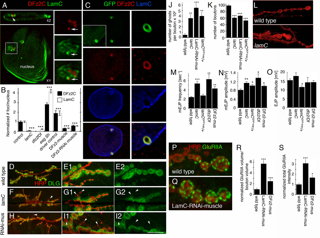
  1. D,E- a wild type NMJ at (A) low and (B) high magnification,

  2. F, G- a lamC null mutant NMJ at (C) low and (D) high magnification, and

  3. H, I- an NMJ from a larva expressing LamC-RNAi in muscles (LamC-RNAi-muscle).

Arrowheads=ghost boutons. Calibration=30µm D, F, H; 12µm E, G, I. J, K- Morphometric analysis of NMJs showing
  1. J- ghost boutons, and

  2. K- bouton number.

N (same order as in graph)= 10, 19, 16, 16. L- Morphology of synaptic boutons in (top) wild type and (bottom) lamC mutant. Calibration=12µm. M–O- Electrophysiological analysis of larval NMJs showing
  1. M- mEJP frequency,

  2. N- mEJPs amplitude, and

  3. O- evoked EJP amplitude.

N (same order as in graph)=8, 8, 5, 5, 7. P,Q- Larval NMJs labeled with HRP and DGluRIIA antibodies in
  1. (P) wild type and

  2. (Q) a larva expressing LamC-RNAi in muscles.

Calibration= 5 µm. R,S- Morphometric analysis of GluRIIA clusters showing
  1. (R) GluRIIA label volume and

  2. (S) total intensity.

  3. ***= p< 0.0001; **= p<0.01; *= p<0.05. Bars in graphs indicate mean±SEM.

Figure 2
Figure 2. Localization of DFz2C/LamC foci at larval muscle nuclei in relationship to DNA, nuclear membrane, nuclear pore complexes, and cytoplasmic 70 KDa-dextran (also see SF1,2)
A–E- DFz2C/LamC foci (arrows) in relationship with
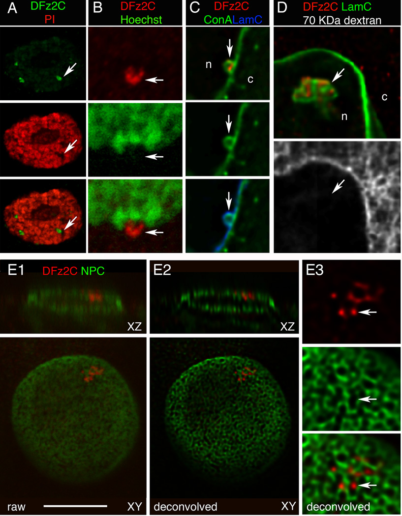
  1. A, B- propidium iodide and Hoechst,

  2. C- the membrane marker Concanavalin-A (ConA; deconvolved),

  3. D- 70 KDa dextran injected into the cytoplasm (deconvolved),

  4. E- mAb414 labeling the nuclear pore complex (NPC;(Davis and Blobel, 1986)).

    1. E1 shows a raw image of the nucleus,

    2. E2 a deconvolution of E1, and

    3. E3 a high magnification view of the focus in E2.
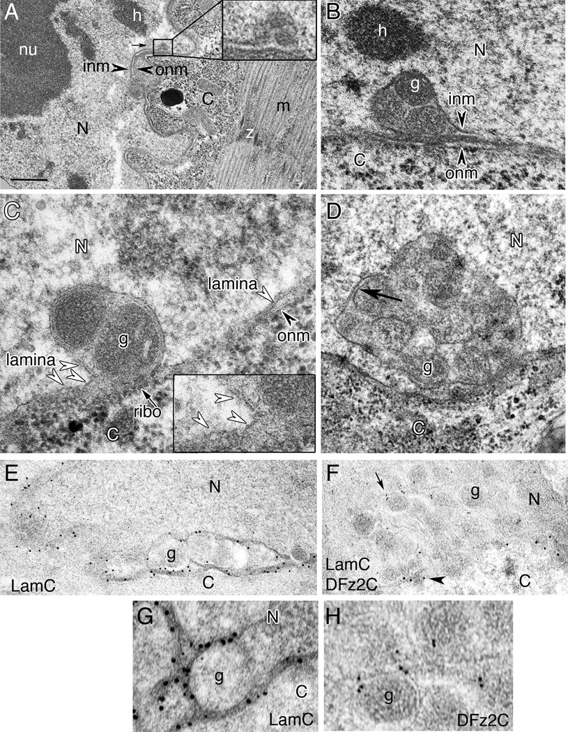
Images are single confocal slices; c= cytoplasm; n=nucleoplasm; Calibration= 14µm A; 8µm B, E1–2; 4µm C, D, E3.
Figure 3
Figure 3. Ultrastructural organization of DFz2C/LamC foci (also see SF3
Transmission electron micrographs of larval muscle or S2 cell nuclei containing DFz2C/LamC foci. A- Low magnification of a focus (box; enlarged in inset) within a muscle nucleus. B–D- High magnification of INM invaginations containing electron dense granules (g) from
  1. B- larval muscle.

  2. C, D- S2 cells. Dense granules appear to be bounded by membrane (arrow in D).

E–H- Immunoelectron micrographs of muscle nuclei labeled with
  1. E, G- anti-LamC and 18nm gold-conjugated second antibody shown at (E) low and (G) high magnification;

  2. F, H- anti-DFz2C and anti-LamC with 12nm and 18nm gold conjugated second antibody, respectively, shown at (F) low and (G) high magnification.
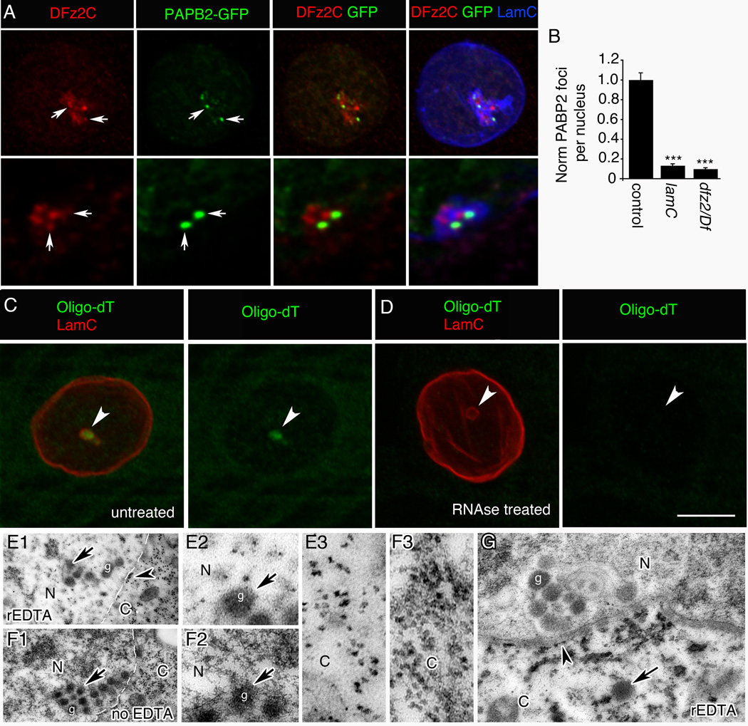
N= nucleus; C= cytoplasm; nu= nucleolus; h=heterochromatin; inm= inner nuclear membrane; onm= outer nuclear membrane; m= myofibrils; z= perforated z band. Calibration=0.5µm A; 0.3µm B, D; 0.4µm E, F; 0.1µm C, G, H.
Figure 4
Figure 4. PABP2 and poly(A) RNA are associated with DFz2C/LamC foci (also see SF4)
A- View of a muscle nucleus showing the relationship between DFz2C/LamC and PABP2-GFP foci (arrows), displayed at (top) low and (bottom) high (deconvolved) magnification. B- PABP2 foci number/muscle nuclei. N (same order as in graph)=229, 175, 301. C, D- Muscle nucleus labeled with poly(dT) FISH and anti-LamC, either
  1. C- without or

  2. D- with RNase treatment.

E–G- View of muscle DFz2/LamC foci in sections
  1. E- treated with rEDTA and

  2. F- not treated with EDTA (same preparation as in (E)). N= nucleus; C= cytoplasm.

    1. E2, F2- High magnification views at the nuclear area of E1 and F1 around a DFz2C granule showing a dark meshwork surrounding the granule, which is bleached after rEDTA treatment (E2), while DFz2C granules retain electron density (F2).

    2. E3, F3- High magnification of E1 and F1, showing ribosomes in the cytoplasm, which retain electron density after EDTA treatment.

  3. G- Low magnification view of a muscle focus showing retention of electron density by DFz2C granules after rEDTA (g; arrow).

  4. Arrowheads=ribosomes at ONM. Arrow=cytoplasmic granule of the same size and morphology as DFz2C granules at INM invaginations.

Calibration= 8µm A (top row); 3µm A (bottom row); 8µm C, D; 0.6µm E1, F1; 0.2µm E2–3, F2–3; 1µm G. ***= p< 0.0001. Bars in graphs indicate mean±SEM.
Figure 5
Figure 5. RNA granules from DFz2C/LamC foci exit larval muscle nuclei
A, B- Nuclei imaged live from larval body wall muscle preparations after incubation with E36 RNA dye. After time-lapse imaging, samples were fixed and labeled with antibodies to DFz2C and/or LamC. N=nucleus; nu=nucleolus. See Supplementary Movie 1 and 2. Calibration=10µm A(top row), B(top row); 4µm A(bottom row), B(bottom row).

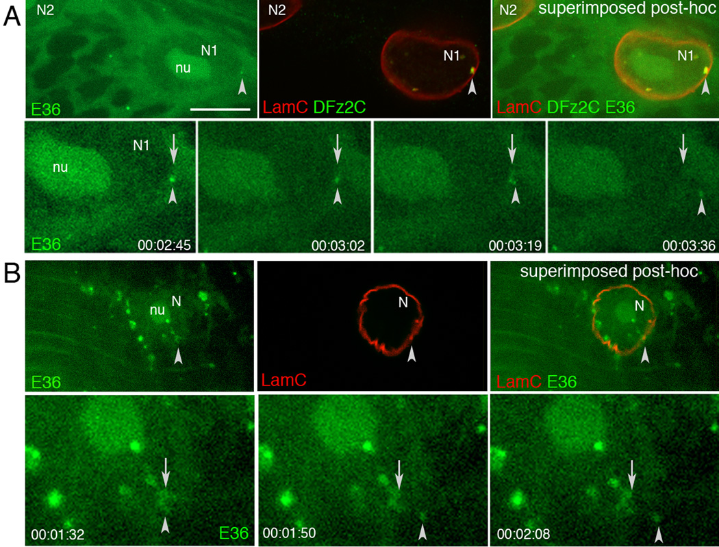
Top rows in A, B show (top panel) a single image of a larval body wall muscle nucleus labeled with E36, (middle panel) the same nucleus after fixation and immunolabeling with DFz2C and/or LamC antibodies, and (right panel) their superposition obtained after resizing using fiduciary markers. Arrowheads=position of the E36 and DFz2C/LamC foci.

Bottom rows in A, B display time-lapse imaging series, showing an E36 labeled granule exiting the nucleus of a larval body wall muscle. Arrows = initial position of the granule; arrowheads= granules while moving away from the nucleus. Time marks=hr:min:sec.

Figure 6
Figure 6. aPKC is required for foci formation (also see SF5)
A–I- Body wall muscle nuclei labeled with antibodies to either Baz, aPKC, PKC-phosphorylated substrate (aPKC subs), or DFz2C, double labeled with antibodies to LamC in wild type and genetic variants altering aPKC activity showing
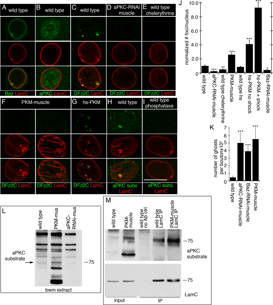
  1. A- localization of Baz at LamC foci,

  2. B- diffuse distribution of aPKC in muscle nuclei,

  3. C- a DFz2C/LamC foci in wild type muscle nucleus,

  4. D- virtual elimination of DFz2C/LamC foci by expressing aPKC-RNAi in muscles,

  5. E- that chelerythrine feeding, decreases DFz2C/LamC foci,

  6. F- that expressing PKM in muscles enlarges and increases LamC foci number, which are devoid of DFz2C,

  7. G- that expressing PKM for just 30 min increases DFz2C/lamC foci number,

  8. H- enrichment of PKC phosphorylated substrates immunoreactivity at LamC foci, and

  9. I- that this label is eliminated after treatment with lambda phosphatase.

Calibration=15µm. J- Foci number per nuclei upon altering aPKC activity. N (same order as in graph)=2146, 532, 726, 677, 713, 601, 595, and 652. K- Ghost bouton number in indicated genotypes. N=6 for each genotype. L- Western blot of body wall muscle extracts probed with anti-P-PKCs, showing a band of the same molecular weight as LamC (arrow) which increases in intensity in PKM-muscle extracts, and which decreases in intensity in aPKC-RNAi-muscle extracts. N=3. M- Immunoprecipitation of body wall muscle extracts with LamC antibody. N=3. ***= p< 0.0001; **= p<0.01; *= p<0.05. Bars in graphs indicate mean±SEM.
Figure 7
Figure 7. par6 transcript is localized to LamC foci and the NMJ, and forms a complex with DFz2C (also see SF5,6)
A–D, G-L In situ hybridization to larval body wall muscles using par6 or wg probes showing
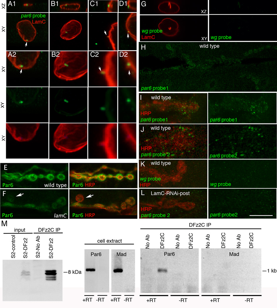
  1. A- association of par6 mRNA with a LamC focus (arrow);

  2. B- localization of par6 transcript near a nuclear membrane fold;

  3. C, D- localization of par6 transcript to cytoplasmically directed projections of the nuclear boundary.

    1. A2–D2 are high magnification views of the nuclei shown in A1–D1.

  4. G- Absence of FISH signal when using a wg probe;

  5. H- a low magnification view of par6 mRNA at the postsynaptic larval NMJ;

  6. I–J synaptic par6 mRNA localization using 2 different par6 probes;

  7. K- absence of synaptic wg mRNA localization;

  8. L- synaptic par6 mRNA localization in a larva expressing LamC-RNAi in muscle, showing virtual elimination of postsynaptic par6 mRNA.

E–F- Larval NMJs labeled with antibodies against HRP and Par6 in
  1. E- wild type and

  2. F- lamC mutant showing a ghost bouton (arrow) devoid of Par6 immunoreactivity and a general decrease in Par6 levels throughout the NMJ.

Calibration =15µm A1, B1, G; 7µm A2, B2; 5µm C1, D1, J; 2.5µm C2, D2; 20µm H; 10µm E, F, I, K, L. M- Immunoprecipitation of par6 RNA using anti-DFz2C. Left: immunoprecipitation of DFz2C fragment using anti-DFz2C. Middle: RT-PCR from S2 cell RNA. Right: RT-PCR of the DFz2C immunoprecipitate.

Comment in

References

    1. Ataman B, Ashley J, Gorczyca D, Gorczyca M, Mathew D, Wichmann C, Sigrist SJ, Budnik V. Nuclear trafficking of Drosophila Frizzled-2 during synapse development requires the PDZ protein dGRIP. Proc Natl Acad Sci U S A. 2006;103:7841–7846. - PMC - PubMed
    1. Ataman B, Ashley J, Gorczyca M, Ramachandran P, Fouquet W, Sigrist SJ, Budnik V. Rapid activity-dependent modifications in synaptic structure and function require bidirectional wnt signaling. Neuron. 2008;57:705–718. - PMC - PubMed
    1. Benoit B, Nemeth A, Aulner N, Kuhn U, Simonelig M, Wahle E, Bourbon HM. The Drosophila poly(A)-binding protein II is ubiquitous throughout Drosophila development and has the same function in mRNA polyadenylation as its bovine homolog in vitro. Nucleic Acids Res. 1999;27:3771–3778. - PMC - PubMed
    1. Bernhard W. A new staining procedure for electron microscopical cytology. J Ultrastruct Res. 1969;27:250–265. - PubMed
    1. Betschinger J, Mechtler K, Knoblich JA. The Par complex directs asymmetric cell division by phosphorylating the cytoskeletal protein Lgl. Nature. 2003;422:326–330. - PubMed

Publication types

MeSH terms