Review
. 2012 Jun;22(3):279-86.
doi: 10.1016/j.sbi.2012.04.005. Epub 2012 May 12.

Molecular recognition and function of riboswitches

Affiliations
Review

Molecular recognition and function of riboswitches

Alexander Serganov et al. Curr Opin Struct Biol. 2012 Jun.

Abstract

Regulatory mRNAs elements termed riboswitches respond to elevated concentrations of cellular metabolites by modulating expression of associated genes. Riboswitches attain their high metabolite selectivity by capitalizing on the intrinsic tertiary structures of their sensor domains. Over the years, riboswitch structure and folding have been amongst the most researched topics in the RNA field. Most recently, novel structures of single-ligand and cooperative double-ligand sensors have broadened our knowledge of architectural and molecular recognition principles exploited by riboswitches. The structural information has been complemented by extensive folding studies, which have provided several important clues on the formation of ligand-competent conformations and mechanisms of ligand discrimination. These studies have greatly improved our understanding of molecular events in riboswitch-mediated gene expression control and provided the molecular basis for intervention into riboswitch-controlled genetic circuits.


Figures

Figure 1
Figure 1
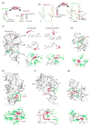
Gene expression control by riboswitches and three-dimensional structures of metabolite-sensing domains (top panels) and their metabolite-binding pockets (bottom panels). The RNA backbone is in a ribbon or stick representation. Ligands are in red stick representation. Green and blue nucleotides use their bases and backbones, respectively, for hydrogen bonding (dashed lines) with ligands. (a) Repression of transcription by the preQ1 riboswitch. In the absence of ligand, mRNA forms an antiterminator hairpin that allows transcription to proceed. In the presence of preQ1 the metabolite sensor adopts a stable metabolite-bound conformation that triggers formation of transcription terminator and premature termination of transcription. Key complementary sequences are colored. (b) Activation of transcription by the gcvT-type glycine riboswitch. Two sensing domains of the riboswitch are arranged in tandem and are followed by a single expression platform. (c) dG riboswitch (PDB ID: 3SKI). Peripheral nucleotides important for dG recognition are in violet. Top, middle and bottom panels in the middle column show involvement of A71 in the loop-loop interactions, (G•U)•G triple formation and dG binding pocket, respectively, from the dG riboswitch, whereas the right column presents corresponding regions from the guanine riboswitch. (d) preQ1-I riboswitch (PDB ID: 2KFC and 3FU2). (e) c-di-GMP-I riboswitch (PDB ID: 3IRW). A water molecule is shown as a red sphere. (f) c-di-GMP-II riboswitch (PDB ID: 3Q3Z). PK stands for pseudoknot. (g) SAH riboswitch (PDB ID: 3NPQ). Double-headed arrow shows a short distance between the ligand sulfur atom and RNA.
Figure 2
Figure 2
Three-dimensional structures of riboswitches that exhibit cooperativity. RNA representation and color codes in panels (a) and (c) are as in Figure 1. (a) Glycine-sensing domain II of the Vibrio cholerae riboswitch (PDB ID: 3OWI). Mg2+ coordination bonds are depicted in green sticks. (b) Secondary structure schematic (top) and structure (middle) of the tandem Fusobacterium nucleatum glycine riboswitch (PDB ID: 3P49). Sensing domains I and II are in light green and orange, respectively, with quartenary interdomain interactions shaded, indicated with double arrows, and highlighted in brighter colors. Bottom: contacts between P1 helix and J3a/3b region. (c) Structure of the THF riboswitch bound to two molecules of folinic acid (FAJ and FAL) (PDB ID: 3SUH).
Figure 3
Figure 3
Ligand-sensing and discrimination by riboswitches. Arrows indicate conformational differences between structures. (a) Discrimination of dG and guanosine (top panels) by the dG riboswitch revealed by superposition of the dG-bound (green and blue) and guanosine-bound (orange) binding pockets (PDB IDs: 3SKI and 3SKZ) [8]. (b) Re-engineering of the adenine riboswitch to discriminate against adenine and interact with ammeline and azacytosine (top panels). Conformational and bonding differences are shown on the adenine-bound (green and blue, PDB ID: 1Y26) and azacytosine-bound (orange, PDB ID: 3LA5) pockets superimposed on the discriminatory pyrimidine 74 [35]. (c) Blocking of the SAM-binding site by A46 (highlighted in bright colors) in the free structure shown by superposition of the SAM-bound (orange, PDB ID: 3IQP) and free (green, PDB ID: 3IQR) pockets [40].
Figure 4
Figure 4
A model of the SAM-I riboswitch folding [56]. In the unfolded state, the riboswitch adopts a Y-shaped conformation. Addition of Mg2+ facilitates P2–P3 stacking, the pseudoknot interactions (PK), and the close juxtaposition of P1 and P3. SAM binding to this preorganized structure stabilizes P1–P4 stacking and rotates P1 along its axis.

Similar articles

Cited by

References

    1. Mironov AS, Gusarov I, Rafikov R, Lopez LE, Shatalin K, Kreneva RA, Perumov DA, Nudler E. Sensing small molecules by nascent RNA: a mechanism to control transcription in bacteria. Cell. 2002;111:747–756. - PubMed
    1. Winkler W, Nahvi A, Breaker RR. Thiamine derivatives bind messenger RNAs directly to regulate bacterial gene expression. Nature. 2002;419:952–956. - PubMed
    1. Baker JL, Sudarsan N, Weinberg Z, Roth A, Stockbridge RB, Breaker RR. Widespread genetic switches and toxicity resistance proteins for fluoride. Science. 2012;335:233–235. - PMC - PubMed
    1. Breaker RR. Prospects for riboswitch discovery and analysis. Molecular cell. 2011;43:867–879. - PMC - PubMed
    1. Serganov A. The long and the short of riboswitches. Curr Opin Struct Biol. 2009;19:251–259. - PMC - PubMed

Publication types

LinkOut - more resources