. 2012 Aug;167(2):152-61.
doi: 10.1016/j.virusres.2012.04.010. Epub 2012 Apr 28.

Genetic characterization of an insect-specific flavivirus isolated from Culex theileri mosquitoes collected in southern Portugal

Affiliations

Genetic characterization of an insect-specific flavivirus isolated from Culex theileri mosquitoes collected in southern Portugal

Ricardo Parreira et al. Virus Res. 2012 Aug.

Abstract

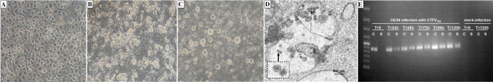
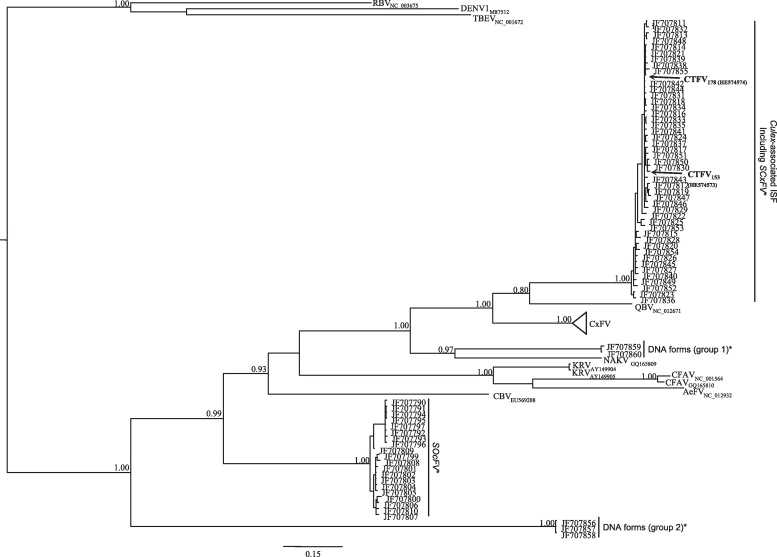
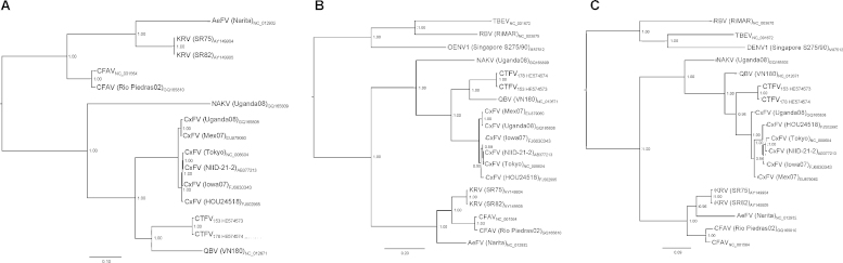
We describe the full genetic characterization of an insect-specific flavivirus (ISF) from Culex theileri (Theobald) mosquitoes collected in Portugal. This represents the first isolation and full characterization of an ISF from Portuguese mosquitoes. The virus, designated CTFV, for Culex theileri flavivirus, was isolated in the C6/36 Stegomyia albopicta (=Aedes albopictus) cell line, and failed to replicate in vertebrate (Vero) cells in common with other ISFs. The CTFV genome encodes a single polyprotein with 3357 residues showing all the features expected for those of flaviviruses. Phylogenetic analyses based on all ISF sequences available to date, place CTFV among Culex-associated flaviviruses, grouping with recently published NS5 partial sequences documented from mosquitoes collected in the Iberian Peninsula, and with Quang Binh virus (isolated in Vietnam) as a close relative. No CTFV sequences were found integrated in their host's genome using a range of specific PCR primers designed to the prM/E, NS3, and NS5 region.


Figures

Fig. 1
Fig. 1
Microscopic observation of C6/36 cells: mock-infected cells (A; 400×), or after infection (day 3) with CTFV strain 153 (B; 400×) or CFAV (C; 200×). (D) Transmission electron micrograph of a thin section of C6/36 cells infected (day 2 post-infection) with CTFV strain 153 (thin arrows) showing multiple round, enveloped viral particles with an electron dense core (thick arrow) accumulated in enlarged cytoplasmic vesicles (scale bar, 200 nm). (E) Kinetics of CTFV153 RNA detection in C6/36 infected and mock-infected cells. At different times after infection, total RNA was extracted from the culture supernatant (S) and cell-sediment (C). After reverse-transcription, a virus specific fragment was amplified with primers SeqC and SeqD (supplementary Table 1). The GeneRuler 1 kb Plus DNA ladder (Fermentas, Vilnius, Lithuania) was used as a molecular weight marker.
Fig. 2
Fig. 2
Bayesian phylogenetic analysis of partial flavivirus NS5 nucleotide sequences. Posterior probability values ≥0.80 are indicated at specific branches. The sequences used are denoted by viral name (Aedes flavivirus – AeFV; Calbertado virus – CBV; cell fusing agent virus – CFAV; Culex flavivirus – CxFV; Culex theileri flavivirus – CTFV; Kamiti River virus – KRV; Nakiwogo virus – NAKV; Quang Binh virus – QBV; Tick-Borne Encephalitis virus – TBEV; Rio Bravo virus – RBV; Dengue virus serotype 1 – DENV1), and accession number. The sequences solely indicated by their accession number and referred with an asterisk [SCxFV (Spanish Culex flavivirus), SOcFV (Spanish Ochlerotatus flavivirus), DNA forms (group 1), and DNA forms (group 2); “DNA forms” indicate these sequences were directly obtained from the amplification of mosquito DNA] were those described by Vázquez et al. (2012). The CTFV sequences here reported are highlighted in bold-face. The size bar indicates 15% of genetic distance.
Fig. 3
Fig. 3
Bayesian phylogenetic analysis of flavivirus ORF (A), NS3 (B) and NS5 (C) amino acid sequences. Posterior probability values ≥0.95 are indicated at specific branches. The sequences used are denoted by viral name (Aedes flavivirus – AeFV; cell fusing agent virus – CFAV; Culex flavivirus – CxFV; Culex theileri flavivirus – CTFV; Kamiti River virus – KRV; Nakiwogo virus – NAKV; Quang Binh virus – QBV; Tick-Borne Encephalitis virus – TBEV; Rio Bravo virus – RBV; Dengue virus serotype 1 – DENV1), viral strain (in parentheses) and accession number. The size bars indicate 10% (A), 20% (B), and 9% (C) of genetic distance.
Fig. 4
Fig. 4
(A) Alignment of conserved GC-rich sequences located in the 3′-UTR of different insect-specific flavivirus (Aedes flavivirus – AeFV; cell fusing agent virus – CFAV; Kamiti River virus – KRV; Culex flavivirus – CxFV; Culex theileri flavivirus – CTFV). The most frequently found nucleotides at each position in the alignment are shaded in gray. (B) Predicted secondary structures for the Culex flavivirus – CxFV (AB262759), cell fusing agent virus (NC_001564) and Culex theileri flavivirus – CTFV (HE574574). SL indicates stem-loop structures. The AUG translation initiation codon is indicated in bold-face. (C) Computer-generated secondary structure analysis of possible interactions between the genomic plus-strand RNA ends of CTFV178 (HE574574). Sections of the 5′- and 3′-UTR are connected by a poly(A) insert (stuffer DNA) simulating most of the viral coding region. The AUG codon is indicated by the arrow. Regions of extended complementarity between both genome ends are boxed. The putative SL2 stem-loop that characterizes the 5′-UTR is indicated by 5′SL2.

Similar articles

Cited by

References

    1. Almeida A.P., Galão R.P., Sousa C.A., Novo M.T., Parreira R., Pinto J., Piedade J., Esteves A. Potential mosquito vectors of arboviruses in Portugal: species, distribution, abundance and West Nile infection. Transactions of the Royal Society of Tropical Medicine and Hygiene. 2008;102:823–832. - PubMed
    1. Almeida A.P., Freitas F.B., Novo M.T., Sousa C.A., Rodrigues J.C., Alves R., Esteves A. Mosquito surveys and West Nile virus screening in two different areas of southern Portugal, 2004–2007. Vector-Borne and Zoonotic Diseases. 2010;10:673–680. - PubMed
    1. Blitvich B.J., Lin M., Dorman K.S., Soto V., Hovav E., Tucker B.J., Staley M., Platt K.B., Bartholomay L.C. Genomic sequence and phylogenetic analysis of Culex flavivirus, an insect-specific flavivirus, isolated from Culex pipiens (Diptera: Culicidae) in Iowa. Journal of Medical Entomology. 2009;46:934–941. - PMC - PubMed
    1. Buckley A., Gaidamovich S., Turchinskaya A., Gould E.A. Monoclonal antibodies identify the NS5 yellow fever virus non-structural protein in the nuclei of infected cells. Journal of General Virology. 1992;73:1125–1130. - PubMed
    1. Bulich R., Aaskov J.G. Nuclear localization of dengue 2 virus core protein detected with monoclonal antibodies. Journal of General Virology. 1992;73:2999–3003. - PubMed

Publication types