Review
. 2013 Feb;63(2):254-66.
doi: 10.1016/j.yhbeh.2012.04.016. Epub 2012 May 2.

Neuroendocrine aspects of catamenial epilepsy

Affiliations
Review

Neuroendocrine aspects of catamenial epilepsy

Doodipala Samba Reddy. Horm Behav. 2013 Feb.

Abstract

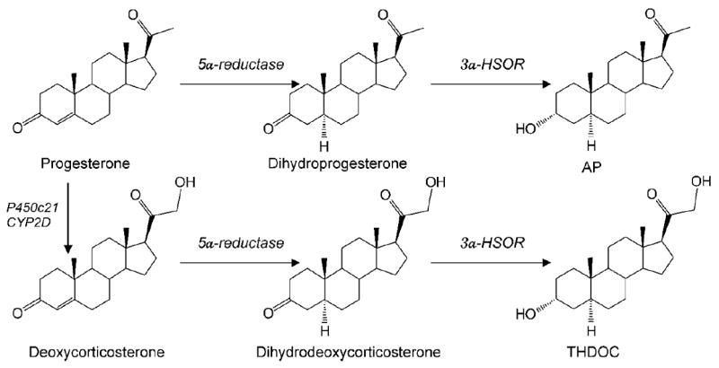
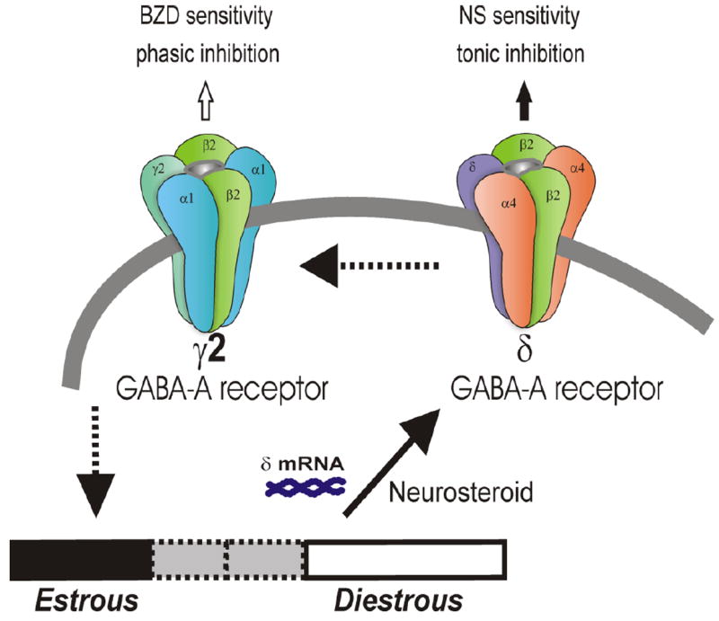
This review describes the neuroendocrinological aspects of catamenial epilepsy, a menstrual cycle-related seizure disorder in women with epilepsy. Catamenial epilepsy is a multifaceted neuroendocrine condition in which seizures are clustered around specific points in the menstrual cycle, most often around perimenstrual or periovulatory period. Three types of catamenial seizures (perimenstrual, periovulatory and inadequate luteal) have been identified. The molecular pathophysiology of catamenial epilepsy remains unclear. Cyclical changes in the circulating levels of estrogens and progesterone (P) play a central role in the development of catamenial epilepsy. Endogenous neurosteroids such as allopregnanolone (AP) and allotetrahydrodeoxycorticosterone (THDOC) that modulate seizure susceptibility could play a critical role in catamenial epilepsy. In addition, plasticity in GABA-A receptor subunits could play a role in the enhanced seizure susceptibility in catamenial epilepsy. P-derived neurosteroids such as AP and THDOC potentiate synaptic GABA-A receptor function and also activate extrasynaptic GABA-A receptors in the hippocampus and thus may represent endogenous regulators of catamenial seizure susceptibility. Experimental studies have shown that neurosteroids confer greater seizure protection in animal models of catamenial epilepsy, especially without evident tolerance to their actions during chronic therapy. In the recently completed NIH-sponsored, placebo controlled phase 3 clinical trial, P therapy proved to be beneficial only in women with perimenstrual catamenial epilepsy but not in non-catamenial subjects. Neurosteroid analogs with favorable profile may be useful in the treatment of catamenial epilepsy.


Conflict of interest statement

Conflict of Interest Statement

The author declares that the research was conducted in the absence of any commercial or financial relationships that could be construed as a potential conflict of interest.

Figures

Fig. 1
Fig. 1. Temporal relationship between ovarian hormones and occurrence of catamenial seizures during the menstrual cycle
The upper panel illustrates the strong relationship between seizure frequency and estradiol/P levels. The lower panel illustrates the three types of catamenial epilepsy. The vertical gray bars (left and right) represent the likely period for the perimenstrual (C1) type, while the vertical gray bar (middle) represents the likely period for the periovulatory (C2) type. The horizontal dark gray bar (bottom) represents the inadequate luteal (C3) type that likely occur starting early ovulatory to menstrual phases. In general, the female reproductive cycle is estimated to last 29 days. Day 1 is the onset of menstruation, and ovulation occurs 14 days before the onset of menstruation. The menstrual cycle is divided into four phases: (i) menstrual phase, days -3 to +3; (ii) follicular phase, days +4 to +9; (iii) ovulatory phase, days +10 to +16; and (iv) luteal phase, days +17 to -4. The early follicular phase is associated with low levels of estrogens and P. The synthesis and secretion of estrogens and P from the ovaries is controlled primarily by the hypothalamic GnRH and pituitary gonadotropins, FSH and LH. As ovulation approaches, the level of estrogen rises and triggers the release of a large surge of LH leading to ovulation. Following ovulation, the ruptured follicle luteinizes and forms a corpus luteum that secretes P and estrogen. Estradiol is secreted in the second half of the follicular phase and increases to a peak at midcycle, while P is elevated during the luteal phase and declines before menstruation begins. The neurosteroid AP is increased in parallel to its precursor, P (Reddy, 2009).
Fig. 2
Fig. 2. Biosynthesis of neurosteroids AP and THDOC in the brain
Enzymatic pathways for the production of two prototype neurosteroids AP and THDOC is illustrated from steroid precursors. P and deoxycorticosterone undergo two sequential A-ring reduction steps catalyzed by 5α-reductase and 3α-HSOR to form the 5α, 3α-reduced neurosteroids. The conversion of P or deoxycorticosterone into neurosteroids occurs in several regions within the brain. The 5α-reductase, 3α-hydroxysteroid oxidoreductase (3α-HSOR), cytochrome P450 21-hydroxylase (P450c21) and other enzymes are present in the brain.
Fig. 3
Fig. 3. Neurosteroid modulation of GABA-A receptors
AP and related neurosteroids binds and potentiate the GABA-A receptor function leading protective effects against seizures. GABA-A receptors are believed to be pentameric with five protein subunits that form the chloride ion channel pore. The AP and THDOC binding site is thought to be at the “neurosteroid binding site”, which is distinct from sites for GABA, benzodiazepines and barbiturates. Postsynaptic GABA-A receptors, which are pentameric chloride channels composed of 2α2βγ subunits, mediate the phasic portion of GABAergic inhibition, while extrasynaptic GABA-A receptors, pentamers composed of 2α2βδ subunits, primarily contribute to tonic inhibition in the hippocampus. Neurosteroids activate both synaptic and extrasynaptic receptors and enhance the phasic and tonic inhibition. Therefore, they may promote maximal protection against seizure susceptibility.
Fig. 4
Fig. 4. A proposed model of ovarian-cycle related changes in the plasticity and function of synaptic and extrasynaptic GABA-A receptors in the hippocampus
Postsynaptic GABA-A receptors, which are pentameric chloride channels composed of 2α2βγ subunits, mediate the phasic portion of GABAergic inhibition, while extrasynaptic GABA-A receptors, pentamers composed of 2α42βδ subunits, primarily contribute to tonic inhibition in the hippocampus. The abundance of δ-subunit-containing GABA-A receptors is elevated at diestrous (high P) than estrous (low P) stage. Such plasticity promotes greater tonic inhibition in the hippocampus and thereby provides resistance against seizure susceptibility (Reddy et al., 2011).

References

    1. Abassi F, Krumholz A, Kittner SJ, Langenberg P. Effects of menopause on seizures in women with epilepsy. Epilepsia. 1999;42:205–210. - PubMed
    1. Agís-Balboa RC, Pinna G, Zhubi A, Maloku E, Veldic M, Costa E, Guidotti A. Characterization of brain neurons that express enzymes mediating neurosteroid biosynthesis. Proc Natl Acad Sci USA. 2006;103:14602–14607. - PMC - PubMed
    1. Amado D, Cavalheiro EA. Hormonal and gestational parameters in female rats submitted to the pilocarpine model of epilepsy. Epilepsy Res. 1998;32:266–274. - PubMed
    1. Bäckström T. Epileptic seizures in women related to plasma estrogen and progesterone during the menstrual cycle. Acta Neurol Scand. 1976;54:321–347. - PubMed
    1. Bäckström T, Zetterlund B, Blom S, Romano M. Effect of intravenous progesterone infusions on the epileptic discharge frequency in women with partial epilepsy. Acta Neurol Scand. 1984;69:240–248. - PubMed

Publication types

MeSH terms