. 2012 Jul 1;53(1):64-71.
doi: 10.1016/j.freeradbiomed.2012.04.020. Epub 2012 May 1.

Systemic administration of the iron chelator deferiprone protects against light-induced photoreceptor degeneration in the mouse retina

Affiliations

Systemic administration of the iron chelator deferiprone protects against light-induced photoreceptor degeneration in the mouse retina

Delu Song et al. Free Radic Biol Med. .

Abstract

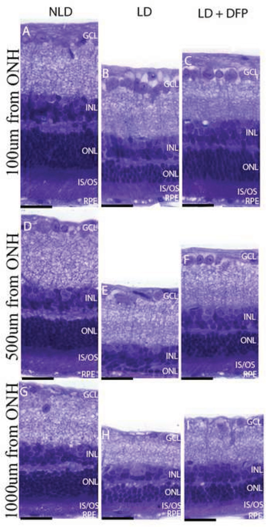
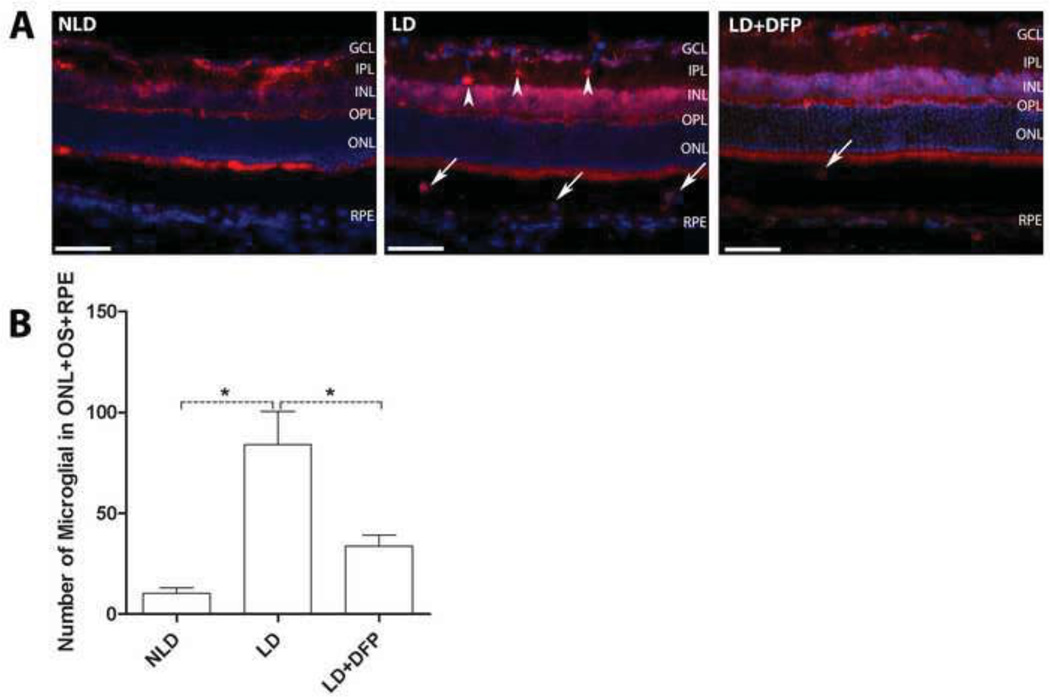
Oxidative stress plays a key role in a light-damage (LD) model of retinal degeneration as well as in age-related macular degeneration (AMD). Since iron can promote oxidative stress, the iron chelator deferiprone (DFP) was tested for protection against light-induced retinal degeneration. To accomplish this, A/J mice were treated with or without oral DFP and then were placed in constant bright white fluorescent light (10,000 lx) for 20 h. Retinas were evaluated at several time points after light exposure. Photoreceptor apoptosis was assessed using the TUNEL assay. Retinal degeneration was assessed by histology 10 days after exposure to damaging white light. Two genes upregulated by oxidative stress, heme oxygenase 1 (Hmox1) and ceruloplasmin (Cp), as well as complement component 3 (C3) were quantified by RT-qPCR. Cryosections were immunolabeled for an oxidative stress marker (nitrotyrosine), a microglial marker (Iba1), as well as both heavy (H) and light (L) ferritin. Light exposure resulted in substantial photoreceptor-specific cell death. Dosing with DFP protected photoreceptors, decreasing the numbers of TUNEL-positive photoreceptors and increasing the number of surviving photoreceptors. The retinal mRNA levels of oxidative stress-related genes and C3 were upregulated following light exposure and diminished by DFP treatment. Immunostaining for nitrotyrosine indicated that DFP reduced the nitrative stress caused by light exposure. Robust H/L-ferritin-containing microglial activation and migration to the outer retina occurred after light exposure and DFP treatment reduced microglial invasion. DFP is protective against light-induced retinal degeneration and has the potential to diminish oxidative stress in the retina.


Conflict of interest statement

Conflict of interest: Dr. Dunaief and ApoPharma have a patent pending on the use of DFP for AMD, and Dr. Dunaief’s lab receives research funding from ApoPharma.

Figures

Fig. 1
Fig. 1
Fluorescence photomicrographs showing TUNEL label in mouse retinas. There are fewer TUNEL-positive photoreceptor nuclei (green, arrow) 72hr following light-damage (LD) in mice treated with DFP compared to retinas from LD mice not treated with DFP (A). Nuclei are counterstained with DAPI (blue). Photoreceptor nuclei reside in the outer nuclear layer (ONL). Scale bars=100µm. (B) Histogram comparing numbers of TUNEL-positive photoreceptor from no light damage (NLD) control (n=3), LD (n=3) and LD+DFP (n=3) mice. The histogram displays the mean (±S.E.M) of total numbers of TUNEL-positive photoreceptors counted in ten sections per retina with an interval of 300µm in the sagittal plane. *Significant difference (P<0.05).
Fig. 2
Fig. 2
Photomicrographs of plastic sections of mouse retinas and plot showing morphologic protection 10 days following LD in LD+DFP retinas. Photomicrographs of plastic sections showing superior retinas of mice in NLD (A), LD (B) and LD+DFP (C) groups. Sagittal plane sections pass through ONH. The photoreceptor nuclei in the ONL and inner/outer segment (IS/OS, labeled with*), are protected from light induced thinning observed in the untreated LD retina. Scale bars=100µm. (D) Plot of the thickness of the ONL, measured in numbers of photoreceptor nuclei per column. Measurements are made in triplicate every 200µm away from the ONH. LD (n=4, red), LD+DFP (n=4, blue) and NLD (n=3, green) retinas 10 days following LD are displayed as mean values (±S.E.M).
Fig. 3
Fig. 3
Photomicrographs of mouse retinas showing protection of DFP against photoreceptor cell loss and inner/outer segment shortening and disruption 10 days following LD. Photomicrographs of 3µm thick plastic section at the indicated distances superior to the ONH from (A, D, G) NLD control, (B, E, H) LD and (C, F, I) LD+DFP retinas 10 days following LD. At 100µm from the ONH (A, B, C), DFP treated retinas have similar numbers of photoreceptor nuclei, compared to NLD, although the IS/OS are shortened. In LD mice without DFP, there is photoreceptor nuclei loss, disorganization and shortening of IS/OS. At 500µm from the ONH (D, E, F), LD retina has marked loss of photoreceptors and IS/OS ablation, but this is diminished by DFP. Light damage is less severe in the periphery. Scale bars=10µm.
Fig. 4
Fig. 4
Graphs showing relative mRNA levels measured by qPCR. DFP decreases oxidative stress and complement markers. Hmox1 and Cp mRNA levels in neural retina (A, B) and Hmox1 mRNA in RPE (D) are significantly upregulated by light exposure; DFP treatment significantly diminishes this upregulation in retina. DFP significantly increases the Rpe65 mRNA level in RPE (E). The increased C3 mRNA level in retina induced by LD is also significantly reduced by DFP treatment (C). LD (n=4), LD+DFP (n=4) and NLD (n=4) retinas are displayed as mean values (±S.E.M). *Significant difference (P<0.05).
Fig. 5
Fig. 5
Comparison of nitrotyrosine levels in retina by immunolabeling and mean pixel density quantification. Monochromatic fluorescence photomicrographs of LD retinas showing stronger nitrotyrosine immunolabeling than DFP treated LD retinas (A). Immunoreactivity was quantified by measuring the mean pixel intensity within the whole retinas. The increased nitrotyrosine immunoreactivity induced by LD is significantly inhibited by DFP treatment (B). Scale bars=100µm. LD (n=3), LD+DFP (n=3) and NLD (n=3) retinas are displayed as mean values (±S.E.M). Significant difference (*P<0.05, **P<0.005).
Fig. 6
Fig. 6
Changes in microglial distribution and number after LD. Fluorescence photomicrographs of Iba1 labeled retinal microglial cells from light-exposed eyes 72hrs following light exposure and counts of these cells in outer retina (ONL+OS+RPE). (A) 72hrs after exposure, labeled microglial were found both in the outer (arrows) and inner (arrow heads) retina. Scale bars=100µm. (B) Quantification of microglial in the outer retina. Compared to NLD control retina, the number of microglia in outer retina increased significantly 72hrs following light exposure, which is significantly diminished by DFP. LD (n=3), LD+DFP (n=3) and NLD (n=3) retinas are displayed as mean values (±S.E.M). *Significant difference (P<0.05).
Fig. 7
Fig. 7
Double-labeling for H/L-ferritin and microglia. Fluorescence photomicrographs for ferritin (green) and microglia (red) in LD retinas: (A, D) Iba1 immunolabeled microglia. (B) H-ferritin immunolabeled cells. (E) L-ferritin immunolabeled cells. (C, F) Merged figures showing co-localization of microglia and ferritins. Scale bars=100µm.

References

    1. A randomized, placebo-controlled, clinical trial of high-dose supplementation with vitamins C and E beta carotene, and zinc for age-related macular degeneration and vision loss: AREDS report no. 8. Arch Ophthalmol. 2001;119:1417–1436. - PMC - PubMed
    1. Taylor HR. Ultraviolet radiation and the eye: an epidemiologic study. Trans Am Ophthalmol Soc. 1989;87:802–853. - PMC - PubMed
    1. Taylor HR, Munoz B, West S, Bressler NM, Bressler SB, Rosenthal FS. Visible light and risk of age-related macular degeneration. Trans Am Ophthalmol Soc. 1990;88:163–173. discussion 173–8. - PMC - PubMed
    1. Marlor RL, Blais BR, Preston FR, Boyden DG. Foveomacular retinitis, an important problem in military medicine: epidemiology. Invest Ophthalmol. 1973;12:5–16. - PubMed
    1. Young RW. Solar radiation and age-related macular degeneration. Surv Ophthalmol. 1988;32:252–269. - PubMed

Publication types

MeSH terms