

Changes in the facial skeleton with aging: implications and clinical applications in facial rejuvenation
- PMID: 22580543
- PMCID: PMC3404279
- DOI: 10.1007/s00266-012-9904-3
Changes in the facial skeleton with aging: implications and clinical applications in facial rejuvenation
Abstract
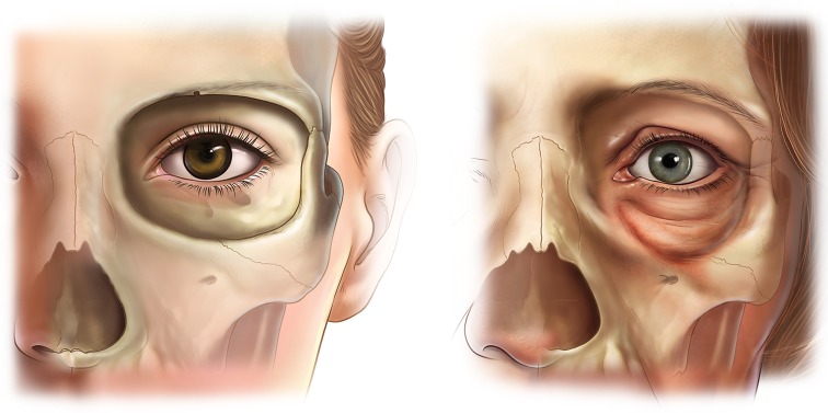
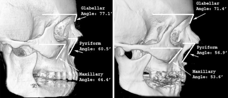
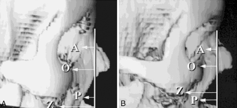
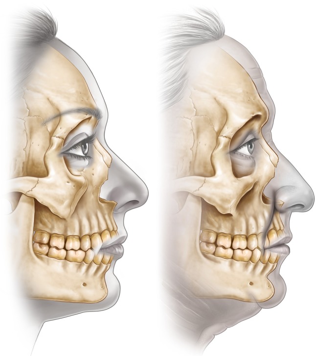
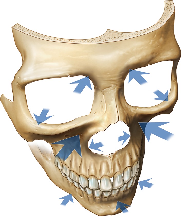
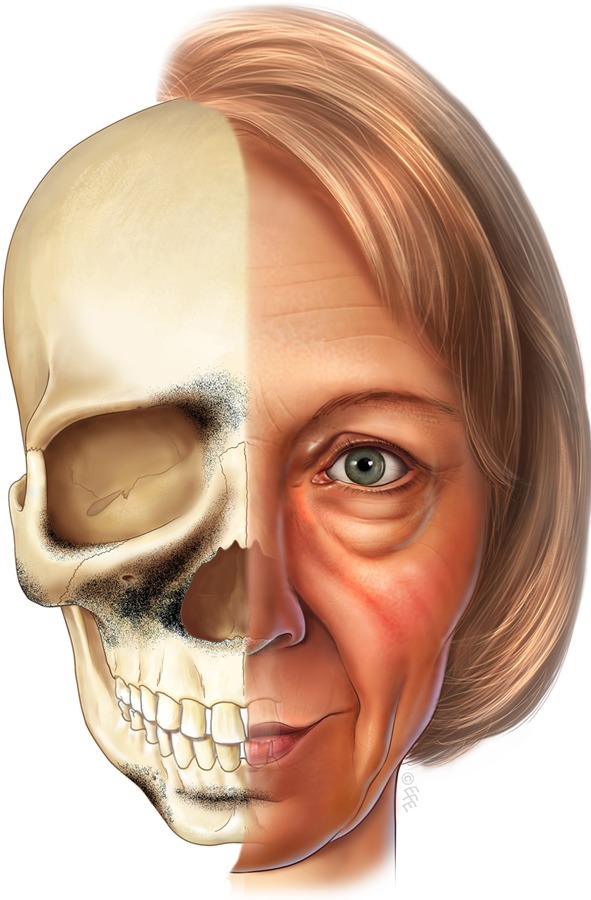
In principle, to achieve the most natural and harmonious rejuvenation of the face, all changes that result from the aging process should be corrected. Traditionally, soft tissue lifting and redraping have constituted the cornerstone of most facial rejuvenation procedures. Changes in the facial skeleton that occur with aging and their impact on facial appearance have not been well appreciated. Accordingly, failure to address changes in the skeletal foundation of the face may limit the potential benefit of any rejuvenation procedure. Correction of the skeletal framework is increasingly viewed as the new frontier in facial rejuvenation. It currently is clear that certain areas of the facial skeleton undergo resorption with aging. Areas with a strong predisposition to resorption include the midface skeleton, particularly the maxilla including the pyriform region of the nose, the superomedial and inferolateral aspects of the orbital rim, and the prejowl area of the mandible. These areas resorb in a specific and predictable manner with aging. The resultant deficiencies of the skeletal foundation contribute to the stigmata of the aging face. In patients with a congenitally weak skeletal structure, the skeleton may be the primary cause for the manifestations of premature aging. These areas should be specifically examined in patients undergoing facial rejuvenation and addressed to obtain superior aesthetic results.
Level of evidence iv: This journal requires that authors assign a level of evidence to each article. For a full description of these Evidence-Based Medicine ratings, please refer to the Table of Contents or the online Instructions to Authors www.springer.com/00266.
Figures
References
-
- Hellman M. Changes in the human face brought about by development. Int J Orthod. 1927;13:475.
-
- Todd TW. Thickness of the white male cranium. Anat Rec. 1924;27:245. doi: 10.1002/ar.1090270504. - DOI
-
- Lasker GW. The age factor in bodily measurements of adult male and female Mexicans. Hum Biol. 1953;25:50. - PubMed
-
- Behrents RG. Growth in the aging craniofacial skeleton. Ann Arbor: University of Michigan Center for Human Growth and Development; 1985.
Publication types
MeSH terms
LinkOut - more resources
Full Text Sources
Other Literature Sources
Medical
Research Materials
