. 2013 Mar 7;32(10):1252-65.
doi: 10.1038/onc.2012.148. Epub 2012 May 14.

Mutant p53 enhances MET trafficking and signalling to drive cell scattering and invasion

Affiliations
Free PMC article

Mutant p53 enhances MET trafficking and signalling to drive cell scattering and invasion

P A J Muller et al. Oncogene. .
Free PMC article

Abstract

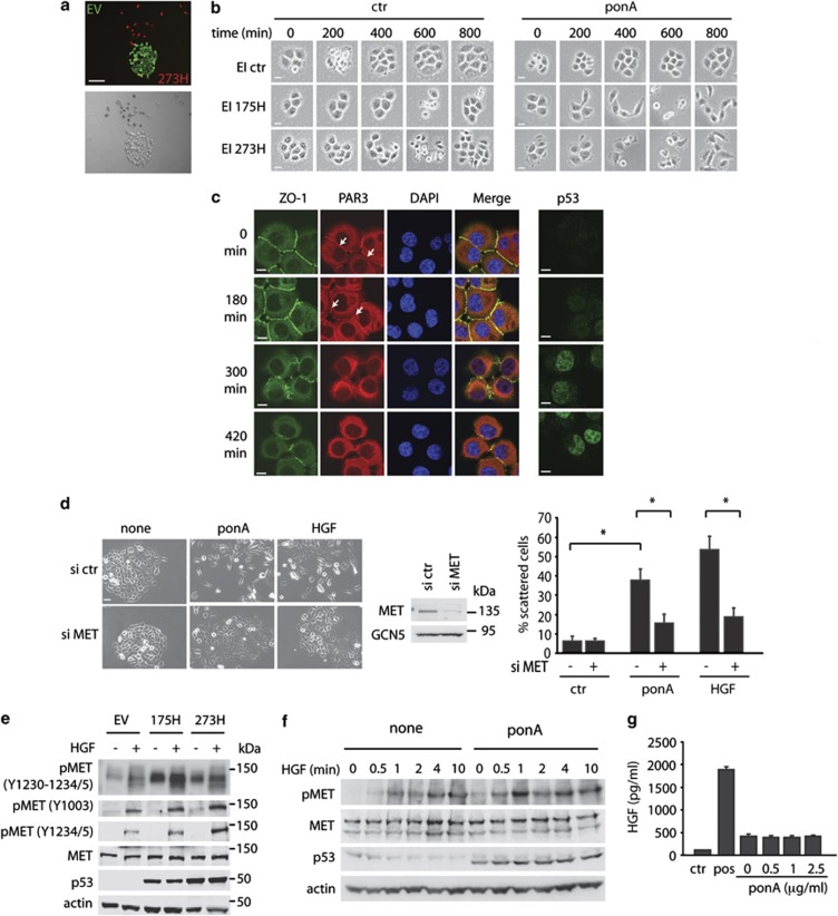
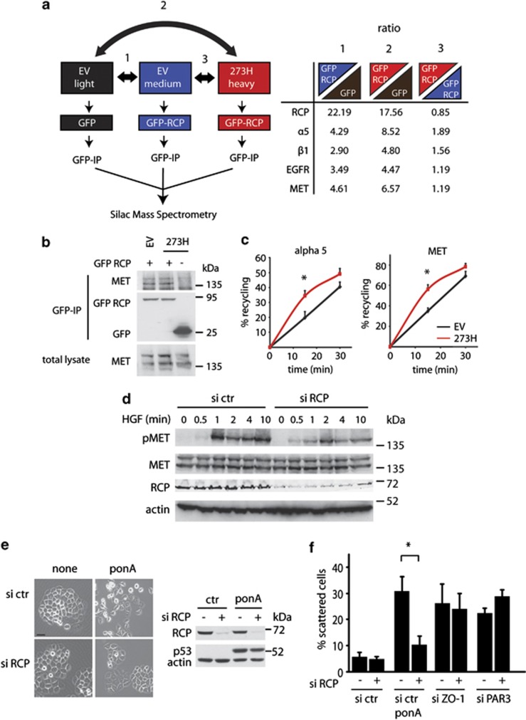
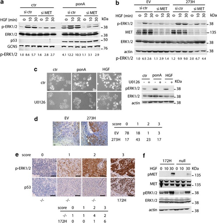
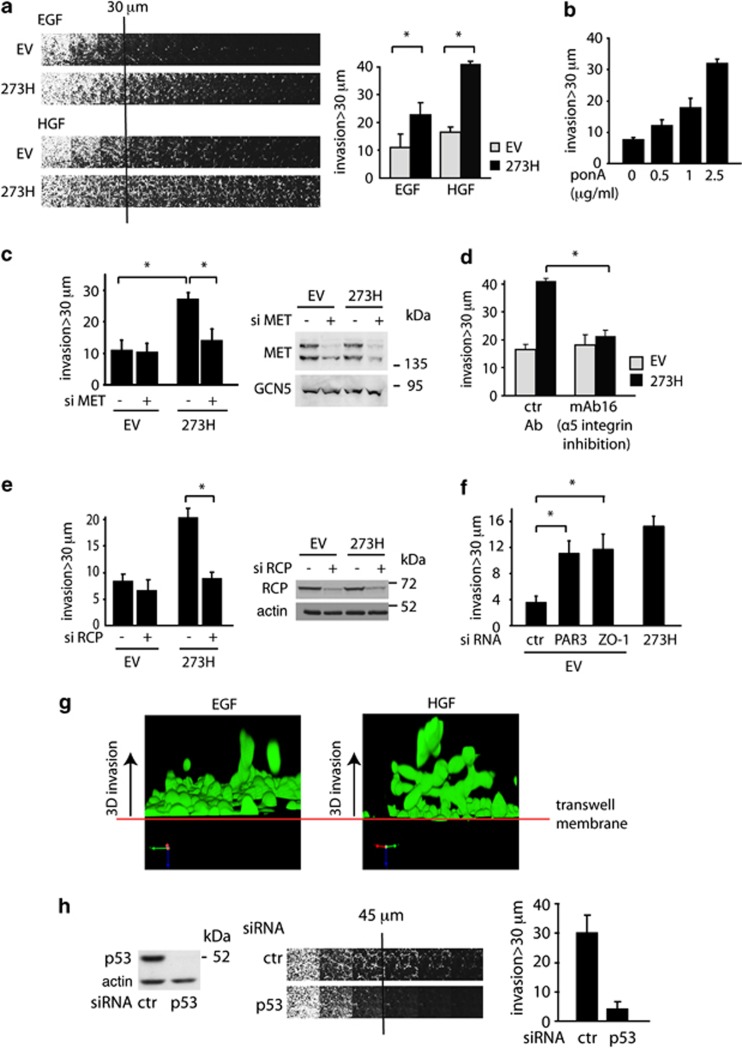
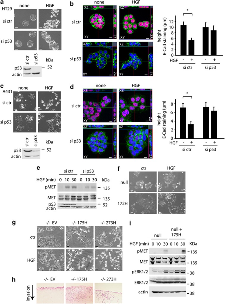
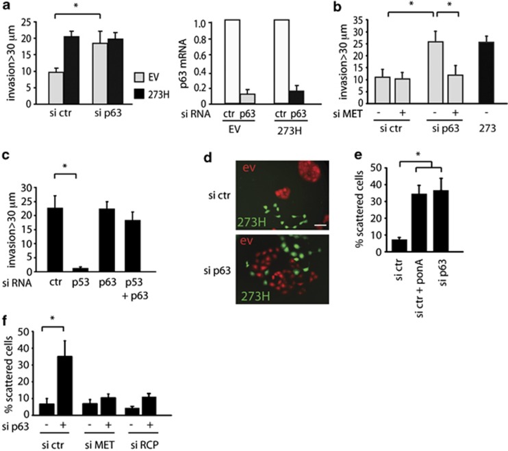
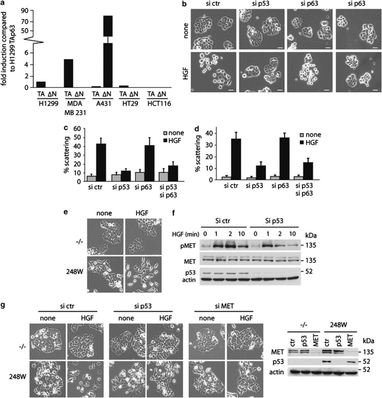
Tumour-derived mutant p53 proteins promote invasion, in part, by enhancing Rab coupling protein (RCP)-dependent receptor recycling. Here we identified MET as an RCP-binding protein and showed that mutant p53 promoted MET recycling. Mutant p53-expressing cells were more sensitive to hepatocyte growth factor, the ligand for MET, leading to enhanced MET signalling, invasion and cell scattering that was dependent on both MET and RCP. In cells expressing the p53 family member TAp63, inhibition of TAp63 also lead to cell scattering and MET-dependent invasion. However, in cells that express very low levels of TAp63, the ability of mutant p53 to promote MET-dependent cell scattering was independent of TAp63. Taken together, our data show that mutant p53 can enhance MET signalling to promote cell scattering and invasion through both TAp63-dependent and -independent mechanisms. MET has a predominant role in metastatic progression and the identification of mechanisms through which mutations in p53 can drive MET signalling may help to identify and direct therapy.


Figures

Figure 1
Figure 1
Mutant p53 enhances MET signalling to drive scattering. (a) Merged red and green channel microscope images and bright field images of control (EV) and mutant p53 (273H) H1299 cells tagged with, respectively, GFP or Cherry. Scale bars indicate 50 μm. (b) EI H1299 cells expressing a vector (ctr) or mutant p53 (175H or 273H) were allowed to form discrete colonies. A total of 2.5 μg/ml ponA was then added to the cells and scattering was monitored using time lapse microscopy. Scale bars indicate 20 μm. (c) Immunostaining for ZO-1, PAR3, DAPI and a merge (left panels) or p53 (right panels) in EI 175H cells after ponA induction. Scale bars indicate 10 μm. Arrows indicate PAR3 staining in the cell–cell junctions. (d) EI 175H cells were transfected with MET siRNA and after 16 h induced with ponA or HGF for 48 h and analysed for scattering (left panels, quantification right panels). Knockdown of MET was verified using immunoblot analysis for MET expression (middle panel) with GCN5 as loading control. Scale bars indicate 50 μm. * indicates statistical significant changes (P<0.05) as determined by a T-test. (e) Mutant p53 (175H or 273H) or control (EV) H1299 cells were analysed for MET expression and MET phosphorylation as assessed by western blot using different pMET antibodies with or without 10 min HGF stimulation. Total MET and actin were used as loading control. (f) Immunoblot analysis showing MET phosphorylation in EI 175H H1299 cells uninduced (none) or induced to express mutant p53 (ponA), incubated with HGF for 0.5, 1, 2, 4 or 10 min. Actin and total MET expression were used as loading controls. (g) ELISA measurement of HGF excretion in the medium of ponA-treated EI 175H cells. Ctr indicates medium that has not been exposed to cells. Pos indicates a positive HGF lysate as included in the ELISA kit.
Figure 2
Figure 2
RCP-dependent recycling of MET contributes to MET signalling in mutant p53 cells. (a) Mutant p53 (273H) or control (EV) H1299 cells were SILAC-labelled with ‘light', ‘medium' or ‘heavy' amino acids and transfected with GFP or GFP-RCP as indicated in the left diagram. Cells were lysed and a co-immunoprecipitation with GFP was used to precipitate RCP-binding proteins. Quantitative mass spectrometry data analysis revealed a ratio of RCP-binding proteins in EV cells and 273H cells compared with GFP-transfected EV cells (comparison 1 and 2), or differential binding to RCP in 273H cells compared with EV cells (comparison 3, right table). (b) MET co-immunoprecipitation with RCP in EV control or 273H H1299 cells that were transfected with GFP or GFP-RCP. Cell lysates were immunoprecipitatedfor GFP and analysed for MET binding as assessed by immunoblot analyses for MET. (c) Recycling assays for alpha 5 integrin and MET in EV and 273H H1299 cells. * indicates statistical significant changes (P<0.05) as determined by a T-test. (d) Immunoblot analysis showing MET phosphorylation in mutant p53 (273H) H1299 that were transfected with siRNA targeting RCP and incubated with HGF for indicated times. Actin and total MET expression were used as loading controls. (e) Scattering of EI 175H cells that were transfected with RCP siRNA and after 16 h induced with ponA (2.5 μg/ml) for 48 h (left panel). Scale bars indicate 50 μm. Knockdown of RCP expression and expression of p53 after ponA (2.5 μg/ml) induction was verified by western blot analysis using actin as a loading control (right panel). (f) Quantification of scattering of EI 175H cells that were transfected with RCP siRNA in combination with or without ZO-1 or PAR3 siRNA and after 16 h induced with ponA for 48 h.
Figure 3
Figure 3
ERK1/2 phosphorylation is required for mutant p53-dependent scattering and invasion. (a) Activation of ERK1/2 in EI 175H cells using phospho-specific ERK1/2 antibodies was determined after induction of mutant p53 with ponA for 24 h. Total ERK1/2 levels are shown as control for equal ERK1/2 expression in all samples and GCN5 was used as loading control. The numbers indicate quantification of pERK1/2 levels as compared with ctr cells transfected with ctr siRNA and not treated with HGF and normalized for total ERK1/2 levels. (b) Activation of ERK1/2 after HGF stimulation in p53 273H and control EV H1299 cells, assessed by western blot using a pERK1/2 antibody. The numbers indicate quantification of pERK1/2 levels as compared with EV cells transfected with ctr siRNA and not treated with HGF and normalized for total ERK1/2 levels. (c) Scattering of EI 175H cells 48 h after ponA, HGF and/or U0126 treatment. Scale bars indicate 50 μm. pERK1/2 expression is shown (right panel) with ERK1/2 and actin as loading controls. (d) Staining of pERK1/2 in xenografts of H1299 EV (8 mice) and 273H cells (7 mice) (left panels). pERK1/2 staining was divided into four intensity categories ranging from 0 for no staining and 3 for the highest intensity. The percentage of staining per category at the invasive edge of the tumour in each section was analysed and the overall percentage of staining for each score is depicted in the table. (e) Immunohistochemical images for pERK1/2 and p53 levels in p53 null and mutant p53 172H (mouse equivalent of human 175H) pancreatic tumours. The scoring indicates the strength of the immunohistochemical signal in which 0 is no staining and 3 is the highest signal. Scale bars indicate 100 μm. The table indicates the numbers of tumours of independent mice in which each score was found. (f) MET and ERK1/2 phosphorylation in cell lines established from the p53 null or mutant p53 (172H) pancreatic tumours after HGF treatment. Total MET, total ERK1/2 and actin were used as loading controls.
Figure 4
Figure 4
Mutant p53 promotes MET-dependent invasion towards HGF. (a) Mutant p53 (273H) or control (EV) H1299 cells were analysed for invasion capacity in fibronectin-supplemented Matrigel using EGF or HGF as a chemo-attractant (left panel). Invasion was quantified as described in the Materials and methods section and values are means±s.e.m. of six replicates from each of three independent experiments (right panel). *indicates statistical significant changes (P<0.05) as determined by a T-test. (b) EI 175H H1299 cells were analysed for invasion capacity in fibronectin-supplemented Matrigel using HGF as a chemo-attractant after induction with increasing doses of ponA. Invasion was quantified as described in the Materials and methods section and values are means±s.e.m. of six replicates from each of three independent experiments (right panel). * indicates statistical significant changes (P<0.05) as determined by a T-test. (c) Mutant p53 (273H) or control (EV) H1299 cells were analysed for invasion after knockdown of MET using HGF as a chemo-attractant. * indicates statistical significant changes (P<0.05) as determined by a T-test. Knockdown of MET was verified by western blot (right panel), using GCN5 as a loading control. (d) Mutant p53 (273H) or control (EV) H1299 cells were analysed for invasion capacity in fibronectin-supplemented Matrigel with monoclonal antibodies against α5 integrin, using HGF as a chemo-attractant. * indicates statistical significant changes (P<0.05) as determined by a T-test. (e) Mutant p53 (273H) or control (EV) H1299 cells were transfected with RCP siRNA and analysed for invasion capacity in fibronectin-supplemented Matrigel using HGF as a chemo-attractant. Knockdown of RCP was verified by western blot using actin as loading control (right panel). * indicates statistical significant changes (P<0.05) as determined by a T-test. (f) H1299 EV cells were transfected with siRNA against ZO-1 or PAR3 and compared with 273H cells for invasion capacity in fibronectin-supplemented Matrigel using HGF as chemo-attractant. * indicates statistical significant changes (P<0.05) as determined by a T-test. (g) 3D reconstructive images of 273H H1299 cells invading towards EGF or HGF. (h) MDA MB231 cells were transfected with siRNA-targeting (mutant) p53 and analysed for invasion capacity into fibronectin-supplemented Matrigel using HGF as chemo-attractant (middle panel). Knockdown was verified by western blot using actin as loading control (left panel). Quantified invasion is shown in the right panel.
Figure 5
Figure 5
Endogenous mutant p53 also promotes scattering. (a and c) Scattering images of HT29 cells (a) or A431 (c) after transfection with control siRNA or p53 siRNA in the presence of 30 ng/ml HGF for 48 h (upper panels). Scale bars indicate 50 μm. Knockdown of p53 was verified by western blot using actin as loading control (bottom panels). (b and d) Ortho-images of Z-stacks of overlay immune fluorescence of HT29 (b) and A431 (d) cells, with views of XY (centre image), XZ (above centre image) or YZ (right of centre image) axes. Cells were transfected with control siRNA or p53 siRNA, then treated or not with 30 ng/ml HGF for 16 h and the height of E-Cadherin staining in cell junctions is shown (right panels). p53 (red), E-cadherin (green), DAPI (blue) and scale bars indicate 10 μm. * indicates statistical significant changes (P<0.05) as determined by a T-test. (e) Immunoblot analysis showing MET phosphorylation in A431 cells transfected with siRNA-targeting (mutant) p53 and incubated with HGF for indicated times. Actin and total MET expression were used as loading controls. (f) Scattering of cell lines established from the p53 null and mutant p53 (172H) pancreatic tumours after HGF treatment for 48 h. Scale bars indicate 50 μm. Scattering (g), invasion in organotypic assays (h) and MET/ ERK1/2 phosphorylation (i) in response to HGF of cell lines established from the p53 null pancreatic tumours that were stably transfected with human mutant p53 175H and 273H. Scale bars indicate 50 μm. Total MET, total ERK1/2 and actin were used as loading controls.
Figure 6
Figure 6
Effect of p63 depletion on cell scattering and invasion. (a) Mutant p53 (273H) or control (EV) H1299 cells were transfected with p63 siRNA and analysed for their invasion capacity to HGF as chemo-attractant. To verify knockdown of p63, relative p63 mRNA expression levels are shown in the right panel. * indicates statistical significant changes (P<0.05) as determined by a T-test. (b) Analysis of invasion of EV cells transfected with a combination of p63 and MET siRNA compared with 273H cells using HGF as chemo-attractant. (c) MDA MB231 cells were analysed for invasion capacity in fibronectin-supplemented Matrigel after knockdown of (mutant) p53 and/or p63, using HGF as chemo-attractant. * indicates statistical significant changes (P<0.05) as determined by a T-test. (d) Fluorescence images of co-cultures of H1299 cells expressing mutant p53 273H and GFP with empty vector (EV) cells expressing Cherry, following transfection of p63 siRNA. Scale bars indicate 50 μm. (e) EI 175H cells were transfected with p63 or ctr siRNA and incubated in ponA (2.5 μg/ml) as indicated. After 48 h, scattering was quantified. * indicates statistical significant changes (P<0.05) as determined by a T-test. Scale bars indicate 50 μm. (f) H1299 cells were transfected with MET or RCP siRNA in combination with p63 siRNA and quantified for scattering 48 h later. * indicates statistical significant changes (P<0.05) as determined by a T-test.
Figure 7
Figure 7
Mutant p53 regulates scattering via p63-dependent and -independent mechanisms. (a) p63 isoform expression in H1299, MDA MB231, A431, HT29 and HCT116 cells using isoform-specific oligos and quantitative RT–PCR. Expression is compared with expression of the TAp63 isoform in H1299 cells and corrected for GAPDH expression in each cell line. (b) Images of scattering of HT29 cells that were transfected with siRNA against (mutant) p53 and/or p63 and after 16 h incubated in HGF for 48 h. (c) Quantification of scattering of HT29 cells that were transfected with siRNA against (mutant) p53 and/or p63 and after 16 h incubated in HGF for 48 h. (d) Quantification of scattering of HT29 cells that were transfected with siRNA against (mutant) p53 and/or p63 for 16 h and incubated in HGF for 48 h. (e) HCT 116 −/− or HCT 248W/− cells were analysed for scattering after HGF treatment for 48 h. (f) HCT116 248W/− cells were transfected with p53 siRNA, serum starved for 1 h and incubated with HGF as indicated. pMET and p53 levels were analysed by western blot, total MET and actin were used as loading control. (g) HCT116 −/− or HCT248 W/− cells were transfected with siRNA against p53 or MET for 16 h and analysed for scattering after HGF treatment for 48 h (left panels). Knockdown of MET and p53 was verified by western blot as indicated in the right panel. Actin was used as loading control.

References

    1. Sigal A, Rotter V. Oncogenic mutations of the p53 tumor suppressor: the demons of the guardian of the genome. Cancer Res. 2000;60:6788–6793. - PubMed
    1. Lang GA, Iwakuma T, Suh YA, Liu G, Rao VA, Parant JM, et al. Gain of function of a p53 hot spot mutation in a mouse model of Li-Fraumeni syndrome. Cell. 2004;119:861–872. - PubMed
    1. Olive KP, Tuveson DA, Ruhe ZC, Yin B, Willis NA, Bronson RT, et al. Mutant p53 gain of function in two mouse models of Li-Fraumeni syndrome. Cell. 2004;119:847–860. - PubMed
    1. O'Farrell TJ, Ghosh P, Dobashi N, Sasaki CY, Longo DL. Comparison of the effect of mutant and wild-type p53 on global gene expression. Cancer Res. 2004;64:8199–8207. - PubMed
    1. Tepper CG, Gregg JP, Shi XB, Vinall RL, Baron CA, Ryan PE, et al. Profiling of gene expression changes caused by p53 gain-of-function mutant alleles in prostate cancer cells. Prostate. 2005;65:375–389. - PubMed

Publication types

MeSH terms