Review
. 2013 Feb 28;32(9):1073-81.
doi: 10.1038/onc.2012.158. Epub 2012 May 14.

MUC1-C oncoprotein as a target in breast cancer: activation of signaling pathways and therapeutic approaches

Affiliations
Review

MUC1-C oncoprotein as a target in breast cancer: activation of signaling pathways and therapeutic approaches

D W Kufe. Oncogene. .

Abstract

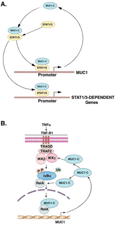
Mucin 1 (MUC1) is a heterodimeric protein formed by two subunits that is aberrantly overexpressed in human breast cancer and other cancers. Historically, much of the early work on MUC1 focused on the shed mucin subunit. However, more recent studies have been directed at the transmembrane MUC1-C-terminal subunit (MUC1-C) that functions as an oncoprotein. MUC1-C interacts with EGFR (epidermal growth factor receptor), ErbB2 and other receptor tyrosine kinases at the cell membrane and contributes to activation of the PI3KAKT and mitogen-activated protein kinase kinase (MEK)extracellular signal-regulated kinase (ERK) pathways. MUC1-C also localizes to the nucleus where it activates the Wnt/β-catenin, signal transducer and activator of transcription (STAT) and NF (nuclear factor)-κB RelA pathways. These findings and the demonstration that MUC1-C is a druggable target have provided the experimental basis for designing agents that block MUC1-C function. Notably, inhibitors of the MUC1-C subunit have been developed that directly block its oncogenic function and induce death of breast cancer cells in vitro and in xenograft models. On the basis of these findings, a first-in-class MUC1-C inhibitor has entered phase I evaluation as a potential agent for the treatment of patients with breast cancers who express this oncoprotein.


Conflict of interest statement

Conflict of Interest: Dr. Kufe has ownership interest in Genus Oncology and is a consultant to the company.

Figures

Figure 1
Figure 1. Structure of the MUC1 heterodimer
A. MUC1 undergoes autoproteolysis at a SEA (sea-urchin sperm protein, enterokinase and agrin) domain to generate two subunits that, in turn, form a stable noncovalent heterodimer. The MUC1-N and MUC1-C nomenclature is used to designate positioning of the subunits after cleavage and to distinguish them from genetic isoforms that are subclassified with Greek characters; for example, ERα and ERβ the PKC isoenzymes, PDGF receptors and others. B. The MUC1-C subunit contains a 58 aa extracellular domain that is glycosylated on asparagine at a N36LT site. The MUC1-C 72 aa cytoplasmic domain interacts with multiple effectors as described below and is sufficient to induce transformation.
Figure 2
Figure 2. Aberrant localization and overexpression of MUC1-C in breast cancer cells
A. The MUC1-N/MUC1-C heterodimer is expressed at the apical border of normal mammary epithelial cells. MUC1-N extends beyond the glycocalyx of the epithelial cells and contributes to the protective mucous barrier. With activation of the epithelial stress response and release of MUC1-N from the cell surface into the mucous gel, MUC1-C functions as a second line of defense to signal stress to the interior of the cell and thereby to protect against loss of integrity of the epithelial layer. Figure modified from Kufe (1). B. In normal epithelial cells, apical MUC1-C is sequestered from receptor tyrosine kinases (RTKs) that localize to the basal-lateral borders. However, with transformation and loss of polarity, MUC1-C is repositioned over the entire cell membrane and forms complexes with RTKs that promote RTK signaling and upregulation of MUC1-C expression. Figure modified from Kufe (1). C. Tissue section containing foci of breast carcinoma cells surrounding normal ducts that was stained with an anti-MUC1 antibody. Expression of MUC1 along the apical borders of normal ductal epithelial cells (arrow) is in contrast to the increased expression of MUC1 over the entire cell membrane and in the cytoplasm of breast cancer cells. Figure originally published in Gaemers et al., J. Biol. Chem. 2001; 276:6191-6199. © the American Society for Biochemistry and Molecular Biology (86).
Figure 3
Figure 3. Interaction between MUC1-C and EGFR at the cell membrane
A. The MUC1-C subunit forms complexes with EGFR at the cell membrane that are mediated extracellularly by galectin-3 bridges (31). The MUC1-C cytoplasmic domain is phosphorylated by EGFR and other RTKs. In turn, the MUC1-C cytoplasmic domain functions as an adaptor for binding of the PI3K SH2 domains and activation of the PI3K→AKT pathway (46; 38). B. The 72 aa MUC1-C cytoplasmic domain is phosphorylated by diverse RTKs and non-receptor tyrosine kinases, providing binding sites for SH2 domains in effectors that, in addition to PI3K (46; 38), include SRC (33), and GRB2 (52).
Figure 4
Figure 4. Intracellular cycling of MUC1-C to the nucleus and mitochondria
MUC1-C cycles from the cell membrane to endosomes and then back to the cell membrane (54; 55). Alternatively, with overexpression in breast cancer cells, MUC1-C accumulates in cytoplasm where it forms homodimers that are mediated by the CQC motif in the MUC1-C cytoplasmic domain (58). MUC1-C dimerization is necessary for transport into the nucleus by importin-β (57). In the nucleus, MUC1-C associates with transcription factors, such as STAT1/3, NF-κB RelA and ERα, and contributes to transactivation of their target genes (1). MUC1-C is also transported to the mitochondrial outer membrane where it blocks activation of the intrinsic apoptotic pathway (59).
Figure 5
Figure 5. MUC1-C activates the Wnt/βcatenin pathway and confers activation of cyclin D1 expression
The MUC1-C cytoplasmic domain contains a SAGNGGSSLS motif that binds directly to the β-catenin Armadillo repeats (insert) (62; 69). GSK3β, an effector of the Wnt pathway, binds to the upstream STDRSPYEKV site, phosphorylates Ser-44 and blocks GSK3β-mediated degradation of β-catenin (63; 69). Phosphorylation of (i) Tyr-46 by EGFR and SRC, and (ii) Thr-41 by PKCδ increases the interaction between MUC1-C and β-catenin (insert) (33; 32; 64). Cytoplasmic MUC1-C homodimers bind to β-catenin and stabilize β-catenin by blocking GSK3β-mediated phosphorylation and degradation of β-catenin. In the nucleus, MUC1-C forms a complex with β-catenin and TCF7L2 on the cyclin D1 promoter and coactivates cyclin D1 expression (73).
Figure 6
Figure 6. MUC1-C forms complexes with the STAT1/3 and NF-κB RelA transcription factors that in turn activate the MUC1 gene in auto-inductive loops
A. Schema for a MUC1-C/STAT1/3 auto-inductive loop involving activation of MUC1 and other STAT1/3-dependent genes. Figure modified from Khodarev et al., 2010 (19). B. Model for the proposed effects of MUC1-C on activation of the NF-κB pathway through interactions with IKKβ-IKKγ and RelA in an auto-inductive loop. Figure modified from Ahmad et al., 2009 (20).

References

    1. Kufe D. Mucins in cancer: function, prognosis and therapy. Nature Reviews Cancer. 2009;9:874–885. - PMC - PubMed
    1. Kufe D, Inghirami G, Abe M, Hayes D, Justi-Wheeler H, Schlom J. Differential reactivity of a novel monoclonal antibody (DF3) with human malignant versus benign breast tumors. Hybridoma. 1984;3:223–232. - PubMed
    1. Ligtenberg MJ, Kruijshaar L, Buijs F, van Meijer M, Litvinov SV, Hilkens J. Cell-associated episialin is a complex containing two proteins derived from a common precursor. J Biol Chem. 1992;267:6171–7. - PubMed
    1. Levitin F, Stern O, Weiss M, Gil-Henn C, Ziv R, Prokocimer Z, et al. The MUC1 SEA module is a self-cleaving domain. J Biol Chem. 2005;280:33374–86. - PubMed
    1. Macao B, Johansson DG, Hansson GC, Hard T. Autoproteolysis coupled to protein folding in the SEA domain of the membrane-bound MUC1 mucin. Nat Struct Mol Biol. 2006;13:71–6. - PubMed

Publication types