

Transcatheter ventricular septal defect (VSD) creation for restrictive VSD in double-outlet right ventricle
- PMID: 22580772
- PMCID: PMC3586406
- DOI: 10.1007/s00246-012-0337-1
Transcatheter ventricular septal defect (VSD) creation for restrictive VSD in double-outlet right ventricle
Abstract
Background: Double-outlet right ventricle (DORV) with a restrictive ventricular septum is a rare but highly morbid phenomenon that can be complicated by progressive left ventricular hypertrophy, arrhythmias, aneurysm formation, severe pulmonary hypertension, and death in the newborn. Surgical creation or enlargement of a ventricular septal defect (VSD) is palliative but may damage the conduction system or the atrioventricular valves in the newborn. This report presents a transcatheter approach to palliation for a newborn that had DORV with a restrictive ventricular septum.
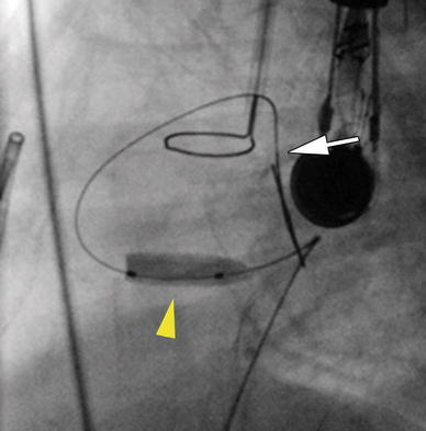
Methods/results: A full-term infant girl (2.9 kg) referred for hypoxia (80% with room air) and murmur was found to have DORV, interrupted inferior vena cava, and restrictive VSD (95-mmHg gradient). Transhepatic access was performed, and an internal mammary (IM) catheter was advanced through the atrial septal defect and into the left ventricle. By transesophageal echocardiographic guidance, a Baylis radiofrequency perforation wire was used to cross the ventricular septum, and the defect was enlarged using a 4-mm cutting balloon. A bare metal stent then was deployed to maintain the newly created VSD. The patient did well after the procedure but required pulmonary artery banding 4 days later. She returned 5 months later with cyanosis and the development of obstructing right ventricle muscle bundles, requiring further surgical palliation.
Conclusions: This report describes the first transcatheter creation of VSD in DORV with a restrictive ventricular septum in a newborn infant. Use of the radiofrequency catheter in combination with cutting balloon dilation and stent implantation is an efficient method for creating a VSD in such a patient.
Figures


References
-
- Edwards JE, James JW, Du SJ. Congenital malformation of the heart: origin of transposed great vessels from the right ventricle associated with atresia of the left ventricular outlet, double orifice of the mitral valve, and single coronary artery. Lab Invest. 1952;1:197–207. - PubMed
-
- Marin-Garcia J, Neches WH, Park SC, Lenox CC, Zuberbuhler JR, Bahnson HT. Double-outlet right ventricle with restrictive ventricular septal defect. J Thorac Cardiovasc Surg. 1978;76:853–858. - PubMed
Publication types
MeSH terms
LinkOut - more resources
Full Text Sources
Medical
