. 2012 Aug;64(8):591-604.
doi: 10.1007/s00251-012-0620-6. Epub 2012 May 13.

Global transcriptional analysis of primitive thymocytes reveals accelerated dynamics of T cell specification in fetal stages

Affiliations

Global transcriptional analysis of primitive thymocytes reveals accelerated dynamics of T cell specification in fetal stages

Nikolai N Belyaev et al. Immunogenetics. 2012 Aug.

Abstract

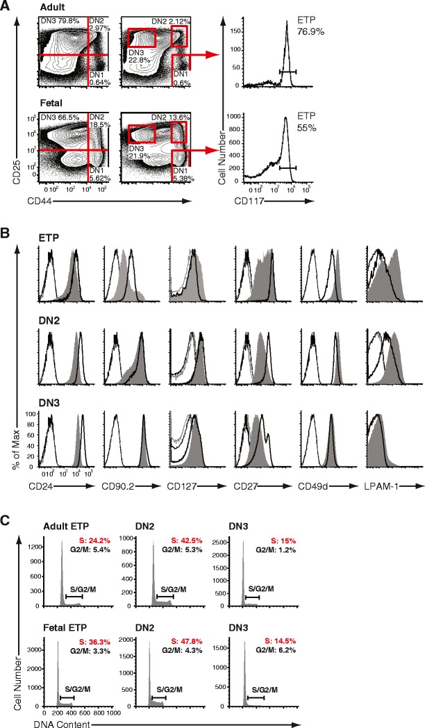
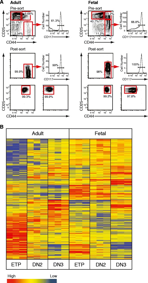
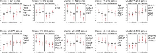
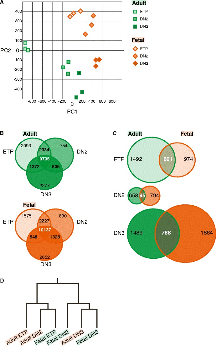
T cell development constitutes a multistage process allowing the dissection of events resulting in cellular commitment and functional specification in a specialized microenvironment. This process is guided by the appropriate expression of regulatory genetic factors like transcriptional activators or repressors which are, in part, dependent on instructive signals of the microenvironment. To date, it remains unclear whether exactly the same genetic mechanism acts in adult compared to fetal T cell development. In order to directly compare T cell commitment during adult and fetal differentiation, we isolated subsequent stages of intrathymic subpopulations starting with early canonical T cell progenitors up to irreversibly committed T cell precursors. The genome-wide analysis revealed several distinct gene clusters with a specific pattern of gene regulation for each subset. The largest cluster contained genes upregulated after transition through the most primitive pool into the next transitory population with a consistently elevated expression of elements associated with ongoing T cell fate specification, like Gata3 and Tcf7, in fetal progenitors. Furthermore, adult and fetal T cell progenitors occupied distinct "transcriptional territories" revealing a precise land map of the progression to final T cell commitment operating in different developmental windows. The presence and/or elevated expression of elements associated with an ongoing establishment of a T cell signature in the most primitive fetal subset is highly suggestive for an extrathymic initiation of T cell specification and underlines the fundamental differences in fetal versus adult lymphopoiesis.


Figures

Fig. 1
Fig. 1
Composition of adult and fetal thymocyte progenitor subsets. a Identification of subsequent stages of early intrathymic differentiation by flow cytometry. Representative histograms illustrating cell surface expression of CD44 and CD25 on lineage negative (double negative, DN) thymocytes in adult (top) and E15.5 fetal thymus (bottom). DN thymocytes were segregated into four populations DN1–4 by expression of CD44 and CD25, as defined by quadrant gating (left panels) or by strictly positioned gates to identify more homogeneous subsets (middle panels). DN1 was CD44+CD25, DN2 was CD44+CD25+, DN3 was CD44lo/intCD25+, and DN4 was CD44lo/intCD25. Early thymic progenitors (ETPs) were defined as CD117+ (receptor-type tyrosine kinase c-Kit) cells in DN1 (right panels). The ETP is a major constituent of adult and fetal DN1. b Representative histograms illustrating expression of CD24, CD90.2, CD127, CD27, CD49d, LPAM-1 on adult (black solid) and E15.5 fetal (gray filled) on DN1 ETP, DN2 and DN3 progenitor thymocytes. Isotype matched controls represent negative staining (adult: faint black, fetal: faint gray lines). c Cell cycle analysis of adult versus fetal thymocyte subsets. Representative histograms illustrating the frequency of cells in S/G2/M in adult (top) and fetal (bottom) DN1 ETP, DN2 and DN3 progenitor thymocytes. DN2 populations contained the highest frequency of cells in cycle, whereas DN3 cells were largely non-dividing
Fig. 2
Fig. 2
Gene expression profiling of adult and fetal double negative thymocytes. a Pre- and post-sort analysis of adult and fetal progenitor thymocytes. Representative histograms illustrating the composition of Lin DN thymocytes from 4-week-old female C57BL/6 mice and E15.5 embryos as resolved by cell surface expression of CD44, CD25 and CD117 before and after FACS purification. Cells were isolated based on CD44 and CD25 in case of DN2 and DN3 (bottom panels). The CD44+CD25 DN1 population was further gated on CD117 (c-Kit) positive cells (middle panels). b Purified ETP, DN2 and DN3 thymocytes from 4–6-week-old female C57BL/6 mice or E15.5 embryos were transcriptionally profiled on the Affymetrix Mouse 430A_2.0 GeneChip. Raw data was transformed and analyzed by Agilent GeneSpring GX 11.0 microarray analysis software. Pearson correlation algorithm generated a heat map illustrating clusters of genes with similar expression levels. Each lane represents an individual technical replicate with pooled material from four (adult) or two (fetal) independently FACS-purified thymic subsets. Individual progenitor populations can be identified by a distinct transcriptional signature
Fig. 3
Fig. 3
Microarray analysis of adult and fetal double negative thymocytes. K-means clustering revealed ten distinct gene clusters with discrete patterns of expression during adult and fetal early intrathymic development
Figure 4
Figure 4
Compartmentalization of adult and fetal progenitor thymocytes according to gene expression signatures. a Principal component analysis based on all regulated genes illustrating “transcriptional territories” of adult and fetal T cell ontogeny. Each progenitor population has a distinct molecular signature based on which novel or aberrant hematopoietic progenitors can be classified. b Venn diagrams illustrating the overlapping and distinctive genetic elements in adult DN1 ETP, DN2 and DN3 progenitors as well as in analogous fetal subpopulations. Filtered sets of genes were grouped according to their absence or presence of transcriptional activity in every subpopulation. The majority of genes both in adult and fetus were expressed during differentiation; however, each population displayed a unique set of transcribed genes. c Venn diagrams illustrating the distribution of genes between adult and fetal DN1 ETP, DN2 and DN3 populations, respectively. Uniquely expressed genes in corresponding populations of adult and fetal development were compared. Analogous populations harbored a set of specific mRNA transcripts that defines each developmental stage. d Self-organizing map of adult and fetal progenitor populations. The dendrogram illustrating relationships between adult and fetal progenitors was derived via a self-organizing map. Fetal DN1 ETP and DN2 progenitors exhibited a closer relationship to the final commitment stage (DN3 stage) as compared to the respective adult populations, suggesting earlier specification of the T cell lineage during fetal ontogeny

References

    1. Akashi K, He X, Chen J, Iwasaki H, Niu C, Steenhard B, Zhang J, Haug J, Li L. Transcriptional accessibility for genes of multiple tissues and hematopoietic lineages is hierarchically controlled during early hematopoiesis. Blood. 2003;101:383–389. doi: 10.1182/blood-2002-06-1780. - DOI - PubMed
    1. Allman D, Sambandam A, Kim S, Miller JP, Pagan A, Well D, Meraz A, Bhandoola A. Thymopoiesis independent of common lymphoid progenitors. Nat Immunol. 2003;4:168–174. doi: 10.1038/ni878. - DOI - PubMed
    1. Belyaev NN, Brown DE, Diaz AI, Rae A, Jarra W, Thompson J, Langhorne J, Potocnik AJ. Induction of an IL7-R(+)c-Kit(hi) myelolymphoid progenitor critically dependent on IFN-gamma signaling during acute malaria. Nat Immunol. 2010;11:477–485. doi: 10.1038/ni.1869. - DOI - PubMed
    1. Benz C, Bleul CC. A multipotent precursor in the thymus maps to the branching point of the T versus B lineage decision. J Exp Med. 2005;202:21–31. doi: 10.1084/jem.20050146. - DOI - PMC - PubMed
    1. Carpenter AC, Bosselut R. Decision checkpoints in the thymus. Nat Immunol. 2010;11:666–673. doi: 10.1038/ni.1887. - DOI - PMC - PubMed