. 2012 May 13;14(6):567-74.
doi: 10.1038/ncb2491.

MYC suppresses cancer metastasis by direct transcriptional silencing of αv and β3 integrin subunits

Affiliations

MYC suppresses cancer metastasis by direct transcriptional silencing of αv and β3 integrin subunits

Hong Liu et al. Nat Cell Biol. .

Abstract

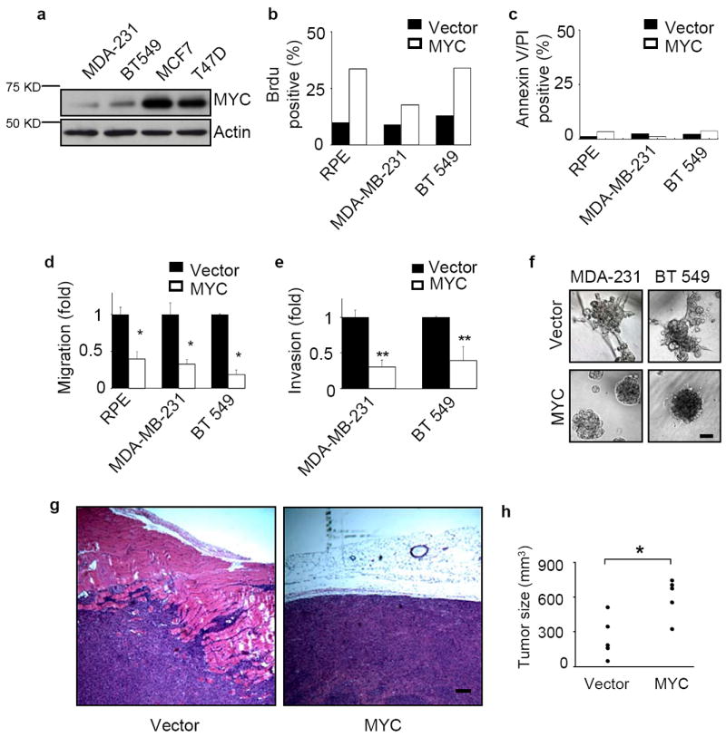
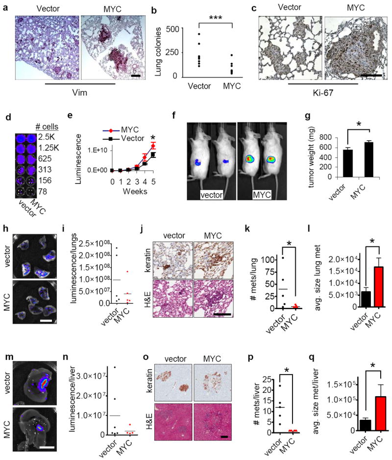
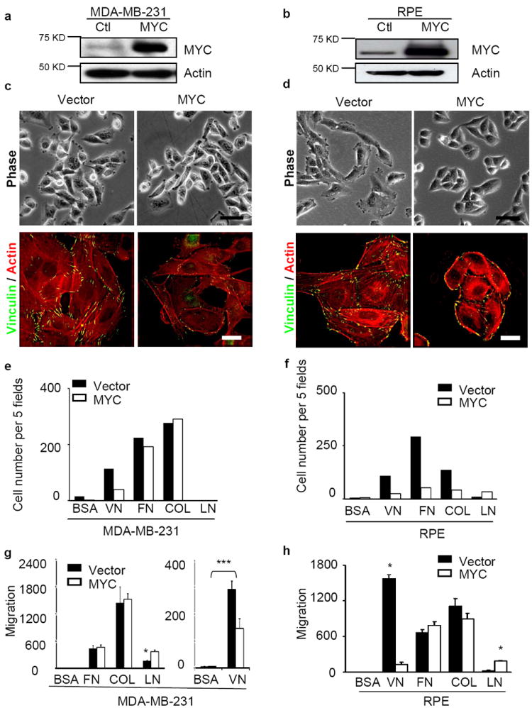
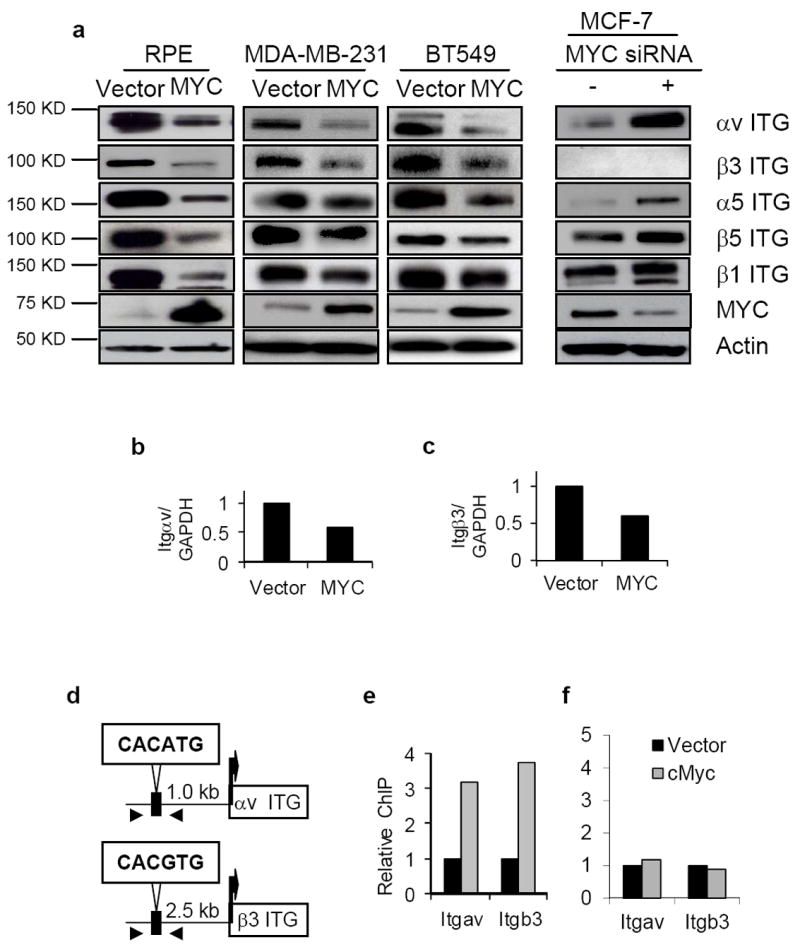
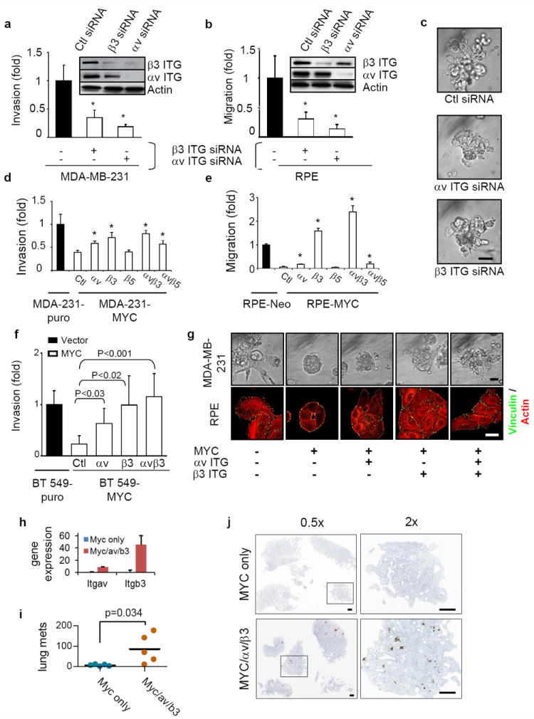
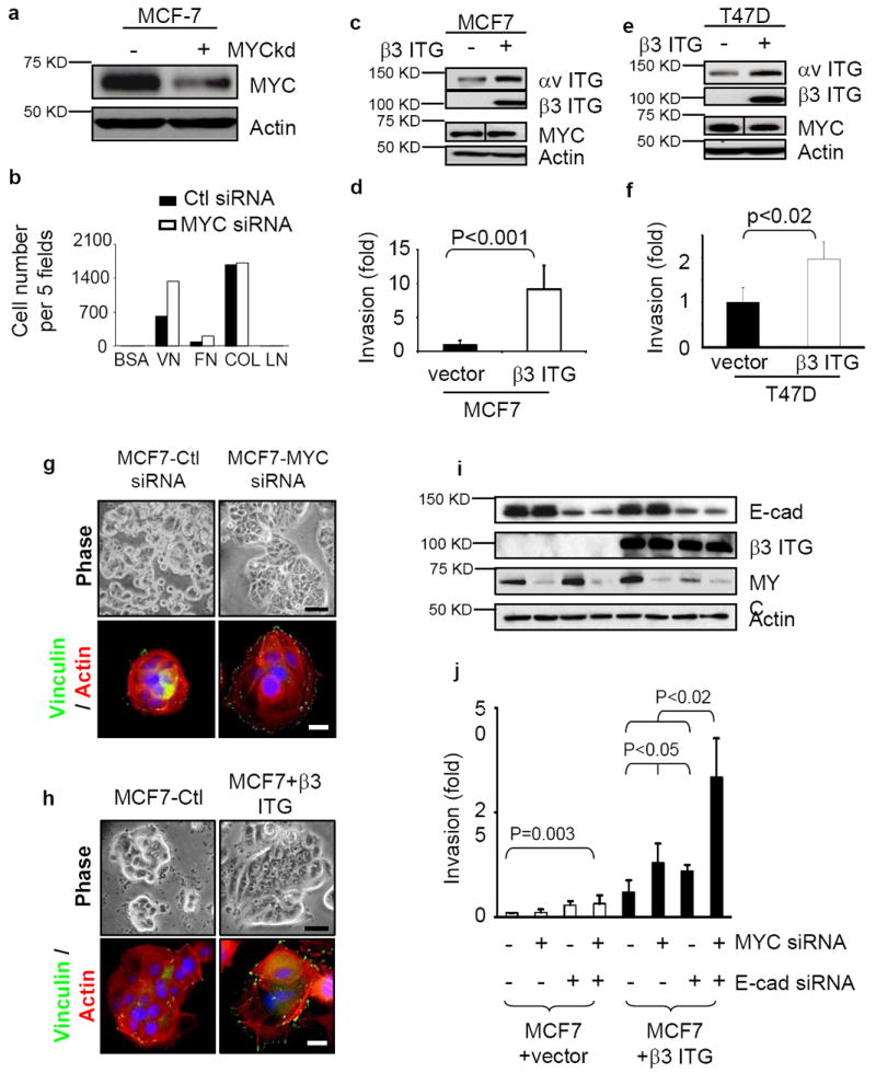
Overexpression of MYC transforms cells in culture, elicits malignant tumours in experimental animals and is found in many human tumours. We now report the paradoxical finding that this powerful oncogene can also act as a suppressor of cell motility, invasiveness and metastasis. Overexpression of MYC stimulated proliferation of breast cancer cells both in culture and in vivo as expected, but inhibited motility and invasiveness in culture, and lung and liver metastases in xenografted tumours. We show further that MYC represses transcription of both subunits of αvβ3 integrin, and that exogenous expression of β3 integrin in human breast cancer cells that do not express this integrin rescues invasiveness and migration when MYC is downregulated. These data uncover an unexpected function of MYC, provide an explanation for the hitherto puzzling literature on the relationship between MYC and metastasis, and reveal a variable that could influence the development of therapies that target MYC.


Conflict of interest statement

COMPETING FINANCIAL INTERESTS

The authors declare no competing financial interests.

Figures

Figure 1
Figure 1
Elevated MYC expression impedes the invasiveness of human breast cancer cells. (a) MYC expression levels in four breast cancer cell lines. The endogenous MYC expression levels of MDA-MB-231, BT549, MCF7, and T47D cells were measured by western blots. (b-c) Ectopic overexpression of MYC increases proliferation but does not affect apoptosis. Cells were labeled with Brdu on tissue culture plastic for 30 minutes at 37°C (b, n=2) or incubated with annexin V and propidium iodide (PI) for 5 minutes at room temperature (c, n=2). Both positive and total nuclei were counted and the results are expressed as mean±SD, * p<0.05. (d) MYC overexpression inhibits cell migration. Migration of MDA-MB-231, BT549 and RPE cells was measured in Boyden-chamber assays (n=3 for each experiment). The cells were transfected with either vector control or a MYC construct. (e) The invasiveness of breast cancer cells is inhibited by MYC overexpression. Cell invasion through Matrigel-coated transwells was measured for MDA-MB-231 (n=3) and BT549 (n=3) cells stably transduced with vector control or exogenous MYC. (f) High level of MYC expression abrogates the invasive phenotype of breast cancer cells grown in 3D Matrigel. Results are shown for day 6. Scale bars: 50 μm. (g-h) MYC overexpression enhances tumor growth but reduces tumor invasion into nearby tissues. MDA-MB-231 cells stably expressing vector or exogenous MYC were inoculated subcutaneously into nude mice. The tumors were collected 4 weeks after injection, sectioned and stained with hematoxilin and eosin (H&E, g) and assessed for size (h, n=5 for each group, p<0.01). Scale bar: 200 μm. Results are expressed as mean± SD. * p<0.05; ** p<0.01.
Figure 2
Figure 2
Elevated MYC expression inhibits metastasis of human breast cancer cells. (a-c) Over-expression of MYC significantly decreases lung metastases of breast cancer cells. MDA-MB-231 cells stably expressing vector or ectopic MYC were injected into nude mice through the tail vein and lung metastases were assessed 6 weeks after injection. Sections of the lungs were stained with antibody against human vimentin (a) and quantification revealed that MYC significantly reduced lung metastasis (b, n=9, p<0.0049). Assessment of Ki-67 (c) revealed increased proliferation in the MYC-expressing cells. Scale bars: 200 μm. (d) Luminescence detection shows comparable luminescence per cell for both vector and MYC over-expressing cells. (e) MYC-expressing cells show more rapid growth at the primary tumor site, assessed by quantitative in vivo imaging (n=5 for each group). (f) Sample images from in vivo imaging of primary tumors for vector and MYC (week 6). (g) Increased size of primary tumors from MYC-expressing cells (week 6; n=5 for each group). (h-i) Decreased metastatic burden in lungs of mice implanted with MYC-expressing cells, assessed by luminescence (week 6; h, sample images, scale bar 1 cm; i, quantification of lung luminescence; n=5 for each group; differences between cells expressing vector alone and those expressing MYC were not statistically significant). (j) Images of lung metastases, stained for human cytokeratins (top), and with H&E (bottom). scale bar=200 μm. (k-l) Quantification of number (k) and size (l) of lung metastases indicates that MYC cells form much fewer metastases but grow to larger size (n=5 for each group). (m-n) Decreased metastatic burden in livers of mice implanted with MYC-expressing cells (week 6; m, sample images, scale bar 1 cm; n, quantification of liver luminescence; differences between conditions were not statistically significant; n=6 for vector, n=5 for MYC). (o) Images of liver metastases, stained for human cytokeratins (top), and with H&E (bottom). Scale bar=200 μm. (q-r) Quantification of number (p) and size (q) of liver metastases indicates that MYC-expressing cells form fewer metastases that grow to larger size (n=5 for each group). Results are expressed as mean± SEM. *, p<0.05; **, p<0.01; ***, p<0.005.
Figure 3
Figure 3
MYC modulates cell shape, actin cytoskeleton, focal adhesion formation, adhesion to and migration towards ECM. (a-b) Overexpression of ectopic MYC in MDA-MB-231 (a) and RPE (b) cells assessed by western blot. (c-d) MYC overexpression inhibits cell spreading, stress fiber and focal adhesion formation. MDA-MB-231 (c) and RPE (d) cells expressing vector or ectopic MYC were cultured for 24 hr, and then stained with anti-vinculin antibody (green) and Texas-Red-conjugated Phalloidin (red). Scale bars: 20 μm for phase contrast and 5 μm for immunofluorescence staining. (e-f) MYC overexpression reduces cell adhesion to ECM. MDA-MB-231 (e) and RPE (f) cells in serum-free medium were plated into 96-well plates coated with purified matrix proteins (VN: vitronectin, FN: fibronectin, Col I: collagen I, LN: laminin; n=2). After 45 minutes, adhered cells were counted in five fields. (g-h) Increased MYC expression blocks cell migration towards vitronectin in a Boyden-chamber assay of MDA-MB-231 (g) and RPE (h) cells (n=3). The results of cell adhesion assay are expressed as mean± SD, *, p<0.05.
Figure 4
Figure 4
MYC down-regulates the expression of αv and β3 integrin genes through binding to their proximal promoters. (a) MYC modulates the abundance of αv and β3 integrins. Total lysates of the indicated cells were fractionated and immunoblotted with the antibodies against the integrins as shown. (b-c) Quantitative PCR assessment of integrin αv (Itgav; b) and β3 (Itgb3; c) expression in response to MYC expression in MDA-MB-231 cells (n=2 for each experiment). (d) Schematic illustration of the E-box motif upstream of αv and β3 integrin genes; arrowheads locate the designed primers for ChIP assay. (e-f) MYC binding to the proximal promoter of αv and β3 integrin genes as determined by ChIP assay. Cross-linked nuclear extracts of MDA-MB-231 transduced with vector only (black columns) or MYC (grey columns) were immunoprecipitated by either anti-MYC antibody or a control IgG. The regions of the MYC binding sites (e) or nonspecific sites (f) upstream of αv or β3 integrin genes were quantified and normalized to IgG pulldown (n=2 for each experiment). Data are expressed as mean± SEM. *, p<0.05; **, p<0.01; ***, p<0.005.
Figure 5
Figure 5
MYC affects breast cancer cell invasiveness by suppressing integrin αv and β3 subunits. (a-b) Knockdown of αv or β3 integrin inhibited invasion (a) and migration (b) in a Boyden-chamber assay, n=3. Knockdown of the integrins by siRNA was confirmed by western blots. (c) Knockdown of αv or β3 integrin inhibits the invasiveness of MDA-MB-231 cells grown in a 3D Matrigel assay for 6 days, n=3. Scale bar: 50μm. (d-e) αv and β3 integrin rescues the compromised migration (d) and invasiveness-(e) elicited by high MYC expression, n=3 for each. MDA-MB-231 and RPE cells overexpressing MYC were transiently transfected with vector, αv, β3, or β5 integrin constructs. Cell invasiveness and migration were assessed by Boyden chamber assay. (f) Inhibition of cancer cell invasiveness by MYC over-expression can be rescued by exogenous expression of αv and β3 integrin subunits in BT549 cells (n=3). (g) Expression of exogenous αv and β3 integrin partially rescued actin cytoskeleton, focal adhesion formation of RPE cells grown on 2D tissue culture plastic dishes, and the compromised invasiveness of MDA-MB-231 cells in a 3D Matrigel assay. RPE or MDA-MB-231 cells, stably expressing the indicated constructs, were plated on cell culture dishes for 24 hours or in 3D Matrigel for 6 days. RPE cells were then stained with anti-vinculin antibody (green) and Texas-Red-conjugated Phalloidin (red). Images of 3D Matrigel culture were obtained by phase contrast microscopy. Scale bars: 50 μm for phase contrast and 5 μm for immunofluorescence staining. (h) Quantitation of Itgav and Itgb3 transcripts by quantitative PCR in MDA/MYC and MDA/MYC/αv/β3 cells, n=3. (i) Quantitation of increased lung metastases in mice orthotopically implanted with MDA/MYC/αvb3 cells as compared to MDA/MYC cells, n=5 for each. (j) Images of lungs of mice orthotopically implanted with MDA/MYC/αvβ3 or MDA/MYC cells. Results are expressed as mean± SD. *, p<0.05.
Figure 6
Figure 6
MYC and E-cadherin can prevent β3 integrin-induced invasion. (a) Suppression of MYC expression by siRNA in MCF7 cells assessed by western blot. (b) Suppression of MYC by siRNA augments cell adhesion to vitronectin and fibronectin. The cells in serum-free medium were plated into 96-well plates coated with purified matrix proteins (VN: vitronectin, FN: fibronectin, Col I: collagen I, LN: laminin). After 45 minutes, adhered cells were counted in five fields, n=2. (c-f) Ectopic expression of β3 integrin in MCF-7 (c) and T47D (e) cells, assessed by Western blot, enhanced invasiveness (d,f; n=3), as assessed by Boyden chamber assay. (g-h) Cell spreading, stress fiber and focal adhesion formation are enhanced in MCF7 cells when MYC is depleted by siRNA (g) or when β3 integrin is exogenously expressed (h). Changes in cell shape, actin cytoskeleton and focal adhesion formation were demonstrated by phase contrast (top) and staining with anti-vinculin (green) and Texas red Phalloidin (bottom). Scale bar: 20 μm for phase contrast and 5 μm for immunofluorescence staining. (i-j) Decreased expression of MYC and E-cadherin by siRNA increased the invasiveness of MCF7 cells only when β3 integrin was expressed. The depletion of MYC and E-cadherin was assessed by western blot (i) and invasiveness was assessed by Boyden chamber assays (j; n=3). Results are expressed as mean± SD.

Comment in

References

    1. Grandori C, Cowley SM, James LP, Eisenman RN. The Myc/Max/Mad network and the transcriptional control of cell behavior. Annu Rev Cell Dev Biol. 2000;16:653–699. - PubMed
    1. Pelengaris S, Khan M, Evan G. c-MYC: more than just a matter of life and death. Nat Rev Cancer. 2002;2:764–776. - PubMed
    1. Dang CV. c-Myc target genes involved in cell growth, apoptosis, and metabolism. Mol Cell Biol. 1999;19:1–11. - PMC - PubMed
    1. Kuttler F, Mai S. c-Myc, Genomic Instability and Disease. Genome Dyn. 2006;1:171–190. - PubMed
    1. Oster SK, Ho CS, Soucie EL, Penn LZ. The myc oncogene: MarvelouslY Complex. Adv Cancer Res. 2002;84:81–154. - PubMed

Publication types

MeSH terms