. 2012 May;29(4):189-98.
doi: 10.1007/s10719-012-9391-4. Epub 2012 May 12.

O-Glycosylation of snails

Affiliations

O-Glycosylation of snails

Herwig Stepan et al. Glycoconj J. 2012 May.

Abstract

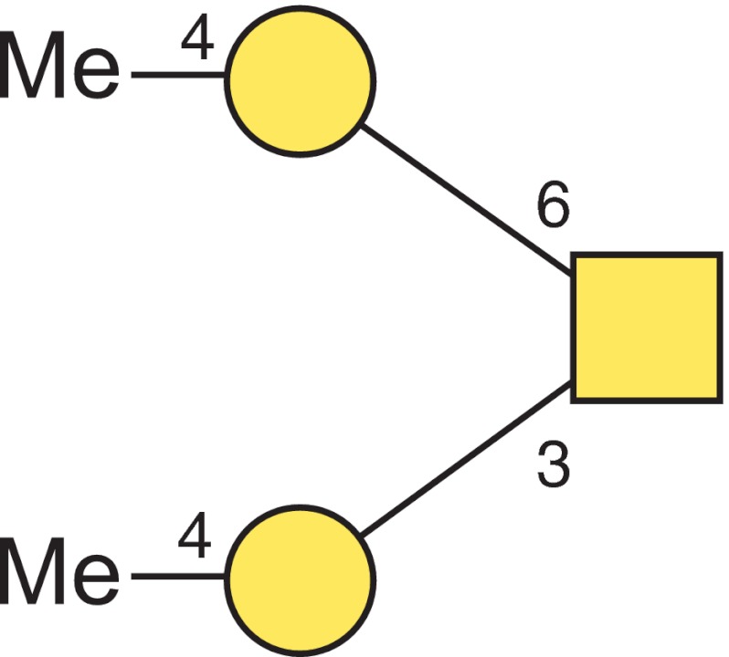
The glycosylation abilities of snails deserve attention, because snail species serve as intermediate hosts in the developmental cycles of some human and cattle parasites. In analogy to many other host-pathogen relations, the glycosylation of snail proteins may likewise contribute to these host-parasite interactions. Here we present an overview on the O-glycan structures of 8 different snails (land and water snails, with or without shell): Arion lusitanicus, Achatina fulica, Biomphalaria glabrata, Cepaea hortensis, Clea helena, Helix pomatia, Limax maximus and Planorbarius corneus. The O-glycans were released from the purified snail proteins by β-elimination. Further analysis was carried out by liquid chromatography coupled to electrospray ionization mass spectrometry and - for the main structures - by gas chromatography/mass spectrometry. Snail O-glycans are built from the four monosaccharide constituents: N-acetylgalactosamine, galactose, mannose and fucose. An additional modification is a methylation of the hexoses. The common trisaccharide core structure was determined in Arion lusitanicus to be N-acetylgalactosamine linked to the protein elongated by two 4-O-methylated galactose residues. Further elongations by methylated and unmethylated galactose and mannose residues and/or fucose are present. The typical snail O-glycan structures are different to those so far described. Similar to snail N-glycan structures they display methylated hexose residues.


Figures

Fig. 1
Fig. 1
Monosaccharide constituent analysis of the core trisaccharide of A. lusitanicus released by [1H] reductive β-elimination. Alditol acetates obtained after acid hydrolysis, reduction with sodium borodeuteride and peracetylation or hydrolysis and peracetylation of already existing alditols were identified by GC-MS. Peaks 1–4 arise from contaminating N-glycans and storage oligosaccharides that could not be completely removed (1: Man, ²H-reduced, 2: Glc, ²H-reduced, 3: Gal, ²H-reduced, 4: GlcNAc, ²H-reduced)
Fig 2
Fig 2
LC-ES-MS/MS spectrum of the core trisaccharide of A. lusitanicus. Registered ions represent proton adducts [M+H]+
Fig 3
Fig 3
Linkage analysis of the core trisaccharide-alditol from A. lusitanicus (a) selected ion chromatogram (m/z 145 and m/z 318) of partially methylated alditol acetates obtained after permethylation with [1H] methyliodide, hydrolysis, reduction with sodium borohydride and peracetylation; (b) EI-MS spectrum and fragmentation pattern of 1,4,5-tri-O-methyl-GalNAc-ol and (c) 2,3,4,6-tetra-O-methyl-galacitol. Characteristic primary selected secondary fragment ions are assigned
Fig 4
Fig 4
EI-MS spectra and fragmentation patterns of (a) 1,4,5-tri-O-deuteromethyl-GalNAc-ol and (b) terminal 4-O-methyl-2,3,6-tri-O-deuteromethyl-galacitol obtained after permethylation of the core trisaccharide alditol from A. lusitanicus with [2H] methyliodide. Characteristic primary and selected secondary fragment ions are assigned
Fig 5
Fig 5
Structure of the core trisaccharide. The structure plot is generated in the notation of the Consortium for Functional Glycomics (http://www.functionalglycomics.org) using the visual editor of “GlycoWorkbench”. This software application is developed and available as part of the EUROCarbDB project (http://www.eurocarb.db.org/applications/ms-tools). Square = GalNAc, Circles = Gal, Me = methyl group
Fig 6
Fig 6
Selected ion chromatograms of partially methylated structures obtained by LC-ESI-MS. a Core trisaccharide carrying in addition one methylated and one unmethylated hexose (m/z 914.4 [M+H]+); b Core trisaccharide with one additional hexose (m/z 738.3 [M+H]+); c Core trisaccharide and its isoform (m/z 576.3 [M+H]+). Square = amino sugar, circle = hexose, Me = methyl group

References

    1. Van den Steen P, Rudd PM, Dwek RA, Opdenakker G. Concepts and principles of O-linked glycosylation. Crit. Rev. Biochem. Mol. Biol. 1998;33:151–208. doi: 10.1080/10409239891204198. - DOI - PubMed
    1. Proszynski TJ, Simons K, Bagnat M. O-glycosylation as a sorting determinant for cell surface delivery in yeast. Mol. Biol. Cell. 2004;15:1533–1543. doi: 10.1091/mbc.E03-07-0511. - DOI - PMC - PubMed
    1. Robledo Y, Marigomez I, Angulo E, Cajaraville MP. Glycosylation and sorting pathways of lysosomal enzymes in mussel digestive cells. Cell. Tissue. Res. 2006;324:319–333. doi: 10.1007/s00441-005-0125-9. - DOI - PubMed
    1. Spiro RG. Protein glycosylation: nature, distribution, enzymatic formation, and disease implications of glycopeptide bonds. Glycobiology. 2002;12:43R–56R. doi: 10.1093/glycob/12.4.43R. - DOI - PubMed
    1. Patsos G, Corfield A. The sugar code. In: Gabius HJ, editor. pp. 111–137. Weinheim: Wiley-VCH; 2009.

Publication types