. 2012 May 13;13(6):612-20.
doi: 10.1038/ni.2305.

DOCK8 functions as an adaptor that links TLR-MyD88 signaling to B cell activation

Affiliations

DOCK8 functions as an adaptor that links TLR-MyD88 signaling to B cell activation

Haifa H Jabara et al. Nat Immunol. .

Erratum in

  • Author Correction: DOCK8 functions as an adaptor that links TLR-MyD88 signaling to B cell activation.
    Jabara HH, McDonald DR, Janssen E, Massaad MJ, Ramesh N, Borzutzky A, Rauter I, Benson H, Schneider L, Baxi S, Recher M, Notarangelo LD, Wakim R, Dbaibo G, Dasouki M, Al-Herz W, Barlan I, Baris S, Kutukculer N, Ochs HD, Plebani A, Kanariou M, Lefranc G, Reisli I, Fitzgerald KA, Golenbock D, Manis J, Keles S, Ceja R, Chatila TA, Geha RS. Jabara HH, et al. Nat Immunol. 2022 May;23(5):815. doi: 10.1038/s41590-022-01180-8. Nat Immunol. 2022. PMID: 35332329 No abstract available.

Abstract

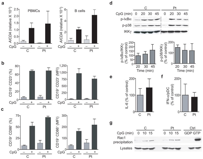
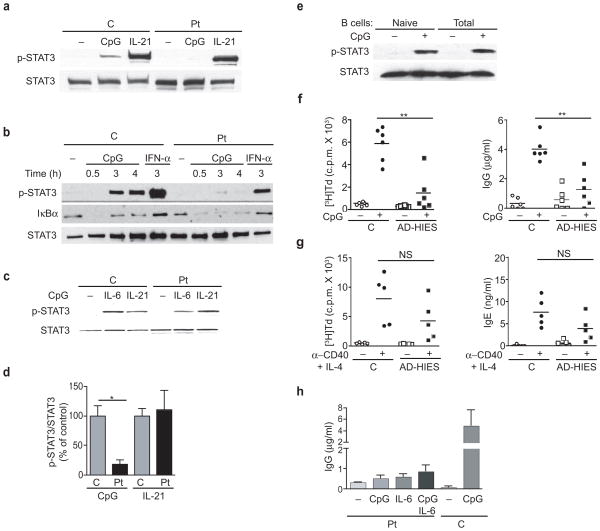
The adaptors DOCK8 and MyD88 have been linked to serological memory. Here we report that DOCK8-deficient patients had impaired antibody responses and considerably fewer CD27(+) memory B cells. B cell proliferation and immunoglobulin production driven by Toll-like receptor 9 (TLR9) were considerably lower in DOCK8-deficient B cells, but those driven by the costimulatory molecule CD40 were not. In contrast, TLR9-driven expression of AICDA (which encodes the cytidine deaminase AID), the immunoglobulin receptor CD23 and the costimulatory molecule CD86 and activation of the transcription factor NF-κB, the kinase p38 and the GTPase Rac1 were intact. DOCK8 associated constitutively with MyD88 and the tyrosine kinase Pyk2 in normal B cells. After ligation of TLR9, DOCK8 became tyrosine-phosphorylated by Pyk2, bound the Src-family kinase Lyn and linked TLR9 to a Src-kinase Syk-transcription factor STAT3 cascade essential for TLR9-driven B cell proliferation and differentiation. Thus, DOCK8 functions as an adaptor in a TLR9-MyD88 signaling pathway in B cells.


Conflict of interest statement

COMPETING FINANCIAL INTERESTS

The authors declare no competing financial interests.

Figures

Figure 1
Figure 1. Impaired antibody responses, failure to maintain serologic memory and decreased memory B cells in DOCK8 deficient patients
(a) Serial antibody titers after re-immunization with TT in two DOCK8 deficient patients aged 8 (Pt. 4) and 15 years (Pt. 7). The dotted line represents the lower limit of the protective antibody titer. (b) Percentage of CD3+ (T) cells and CD19+ (B) cells in PBMCs from DOCK8 deficient patients (Pt) and controls (C (c) Representative flow cytometry analysis of CD19 and CD27 expression by PBMCs from DOCK8 deficient patients and controls. (d) Percentage of CD27+ memory B cells and CD27 naïve B cells in the CD19+ B cell population of DOCK8 deficient patients and controls. Each symbol (b,d) represents an individual subject; small horizontal lines indicate the mean. *P<0.05 and **P<0.001 (Student’s t-test).
Figure 2
Figure 2. Impaired CpG driven B cell proliferation and immunoglobulin (Ig) production to CpG in DOCK8 deficient patients
(a–c) Proliferation and Ig secretion by PBMCs from DOCK8 deficient patients (Pt) and controls (C) in response to stimulation with CpG (a), anti-CD40 + IL-21 (b) and anti-CD40 + IL-4 (c). Not all 10 patients could be studied in each assay. (d) Proliferation, and secretion of IgM and IgG by highly purified total and naïve B cells from controls (n=3 each) and purified total B cells from DOCK8 deficient patients (n=2 each) in response to stimulation with CpG (mean and s.e.). Each symbol (a,b,c) represents an individual subject; small horizontal lines indicate the mean. *P<0.05, and **P<0.001 (Student’s t-test).
Figure 3
Figure 3. Normal CpG-driven upregulation of AICDA, CD23 and CD86 expression, activation of NFκB, p38 MAP kinase and Rac1, and IL-6 secretion in B cells from DOCK8 deficient patients
(a) CpG-induced expression of mRNA for AICDA in PBMCs (left) and purified B cells (right) from patients and controls (n= 2 each). (b,c) Percentage of CD19+ B cells expressing surface CD23 and CD86 (left), and their MFI (right) before and after CpG stimulation of PBMCs from patients and controls (n=3 each). (d) Representative (top) and quantitation (expressed in lower panels as percent of control, n=3 each) of CpG-driven phosphorylation of IκBα and p38 in PBMCs from patients and controls. IKKγ was used as loading control. (e) CpG-driven IL-6 secretion by EBV-B cells from patients relative to controls (n=3 each, mean for controls 55 pg/ml). (f) IFN-α secretion by pDCs in PBMCs from patients and controls in response to stimulation with CpG-A ODN 2216 (n=3 each, mean for controls 22.4 pg/100 pDCs). The number of pDCs/culture was calculated from the percentage of CD123+BDCA-4+ cells in each sample. (g) CpG-driven Rac1 activation in EBV-B cells determined by a GST-PAK precipitation assay followed by immunoblotting for Rac1. Equal volume aliquots of the lysates used in the pull-down assay were immunoblotted for Rac1 as loading controls. Similar data was obtained on three different patients and controls in g; (mean and s.e. in a–f).
Figure 4
Figure 4. CpG-induced DOCK8 dependent STAT3 phosphorylation in B cells is essential for B cell proliferation and IgG production
(a,b) CpG-driven STAT3 Y705 phosphorylation in PBMCs (a) and purified B cells (b) from DOCK8 deficient patients and controls. (c) IL-6 and IL-21 driven STAT3 Y705 phosphorylation in B cells from DOCK8 deficient patients. (d) Quantitation of CpG- and IL-21-driven STAT3 Y705 phosphorylation in B cells from DOCK8 deficient patients and controls (n=3 each). pSTAT3 and STAT3 bands were scanned by densitometry and the pSTAT3/STAT3 ratio was calculated and expressed relative to the mean pSTAT3/STAT3 ratio in controls, which was arbitrarily set at 100% (n=3 per group). (e) CpG-driven STAT3 phosphorylation in total B cells and naïve B cells purified from the same donor. (f,g) CpG-driven proliferation and Ig secretion (n=6 each) (f), and CD40/IL-4 driven proliferation and IgE secretion (n=5 each) (g), by PBMCs from STAT3 deficient AD-HIES patients and controls. (h) Effect of IL-6 (2 ng/ml) on CpG driven IgG secretion by PBMCs from DOCK8 deficient patients (n=3). Results are representative of three patients and three controls in a and b, and three controls in e; mean and s.e. (d, h) and small horizontal lines indicate the mean (f, g). NS, not significant; *P<0.01 and **P<0.001 (Student’s t-test).
Figure 5
Figure 5. CpG-driven phosphorylation of STAT3 in B cells is dependent on Syk, and requires TLR9 and MyD88
(a,b) Syk phosphorylation following CpG stimulation (a) and anti-IgM stimulation (b) of normal and DOCK8 deficient PBMCs detected by immunoblotting with anti-pY352Syk. (c) Quantitation of CpG-driven and BCR-driven Syk Y352 phosphorylation in PBMCs from DOCK8 deficient patients and controls. pSyk and BLNK bands were scanned by densitometry and the pSyk/BLNK ratio was calculated and expressed relative to the mean pSyk/BLNK ratio in controls, which was arbitrarily set at 100% (n=3 per group). (d) Effect of the Syk inhibitor SYKINH-61 on CpG-driven STAT3 Y705 phosphorylation in normal PBMCs. IL-21 driven STAT3 phosphorylation was used as a control. Results are representative of three independent experiments. (e,f) Effect of SYKINH-61 on CpG-driven and CD40 + IL21-driven proliferation (e), IgG production (f) by normal PBMCs (n=4). (g,h) CpG-driven Syk Y352 and STAT3 Y705 phosphorylation in splenic B cells from Tlr9−/− mice and C57BL/6 WT controls (g), and from Myd88−/− mice and BALB/c WT controls (h). Mean and s.e. (c, e–f); NS, not significant; *P<0.05, **P<0.01, and ***P<0.001 (Student’s t-test).
Figure 6
Figure 6. DOCK8 mediates CpG activation of a Pyk2-Src-Syk-STAT3 cascade essential for B cell proliferation and differentiation
(a,b) Representative immunoblot analysis of lysates from CpG-stimulated PBMCs from a DOCK8 deficient patient and a normal control for tyrosine phophorylated proteins (a) and for phosphorylation of Pyk2 at Y402 and Src at Y416 (b). BLNK was used as loading control. The higher amounts of BLNK in PBMCs from the patients reflect the higher percentage of CD19+ B cells in the patients compared to controls (23% versus 7 % in a and 19% versus 8% in b respectively). (c) Immunoblot analysis of lysates from CpG-stimulated splenic B cells from Myd88−/− mice and BALB/c WT controls for pY402Pyk2 and pY416Src. (d–g) Effect of the Pyk2 inhibitor tyrphostin A9 (Tyr A9) and the Src kinase inhibitor PP2 on CpG-driven phosphorylation of Pyk2 at Y402 (d), Src at Y416 (e), Syk Y352 (f), and STAT3 at Y705 (g) in normal PBMCs. (h) Effect of Tyr A9 and PP2 on CpG-driven IgG secretion by normal PBMCs. Stimulation with anti-CD40+IL-21 was used as control. Results in (a–g) are representative of three independent experiments. Data in (h) represent mean and s.e. of three experiments. *P<0.05 and **P<0.01 (Student’s t-test).
Figure 7
Figure 7. DOCK8 associates with MyD88 and Pyk2 and undergoes Pyk2-mediated tyrosine phosphorylation and association with Src/Lyn following TLR9 ligation
(a,b) Association of DOCK8 with MyD88 in 293T cell transfectants (a) and in EBV-B cells (b). (c,d) Association of Pyk2 and MyD88 in EBV-B cells from control (C) and DOCK8 deficient (def) patient (c) and of Pyk2 and DOCK8 EBV-B cells from control (C) and MyD88 def patient (d). Results in a–d are representative of three experiments. (e) DOCK8 tyrosine phosphorylation and association with Src following CpG stimulation of normal B cells. DOCK8 immunoprecipates were probed with anti-p-Tyr, anti-Src and anti-DOCK8 as loading control. Equal volumes of lysates were probed for Src and DOCK8. (f) CpG- driven Lyn phosphorylation in normal PBMCs. Lyn immunopreciptates were probed with anti-pY416Src and with anti-Lyn as loading control. (g) Lyn recruitment to DOCK8 following CpG stimulation of normal PBMCs with CpG. DOCK8 immunoprecipitates were probed with anti-Lyn and anti-DOCK8 as loading control. Upper panel: representative experiment. Lower panel: quantitative analysis of the lyn/DOCK8 band intensity ratio (n=3, mean and s.e.).

References

    1. Yoshida T, et al. Memory B and memory plasma cells. Immunol Rev. 2010;237:117–139. - PubMed
    1. Huggins J, et al. CpG DNA activation and plasma-cell differentiation of CD27- naive human B cells. Blood. 2007;109:1611–1619. - PMC - PubMed
    1. Jiang W, et al. TLR9 stimulation drives naive B cells to proliferate and to attain enhanced antigen presenting function. Eur J Immunol. 2007;37:2205–2213. - PubMed
    1. Giordani L, et al. IFN-alpha amplifies human naive B cell TLR-9-mediated activation and Ig production. J Leukoc Biol. 2009;86:261–271. - PubMed
    1. Carpenter EL, Mick R, Ruter J, Vonderheide RH. Activation of human B cells by the agonist CD40 antibody CP-870,893 and augmentation with simultaneous toll-like receptor 9 stimulation. J Transl Med. 2009;7:93. - PMC - PubMed

Publication types

MeSH terms