. 2012 Jun;18(6):967-73.
doi: 10.1038/nm.2759.

ApoB-containing lipoproteins regulate angiogenesis by modulating expression of VEGF receptor 1

Affiliations

ApoB-containing lipoproteins regulate angiogenesis by modulating expression of VEGF receptor 1

Inbal Avraham-Davidi et al. Nat Med. 2012 Jun.

Abstract

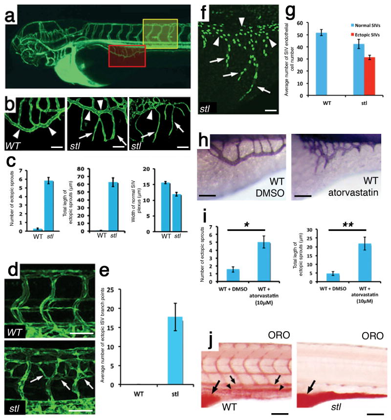
Despite the clear major contribution of hyperlipidemia to the prevalence of cardiovascular disease in the developed world, the direct effects of lipoproteins on endothelial cells have remained obscure and are under debate. Here we report a previously uncharacterized mechanism of vessel growth modulation by lipoprotein availability. Using a genetic screen for vascular defects in zebrafish, we initially identified a mutation, stalactite (stl), in the gene encoding microsomal triglyceride transfer protein (mtp), which is involved in the biosynthesis of apolipoprotein B (ApoB)-containing lipoproteins. By manipulating lipoprotein concentrations in zebrafish, we found that ApoB negatively regulates angiogenesis and that it is the ApoB protein particle, rather than lipid moieties within ApoB-containing lipoproteins, that is primarily responsible for this effect. Mechanistically, we identified downregulation of vascular endothelial growth factor receptor 1 (VEGFR1), which acts as a decoy receptor for VEGF, as a key mediator of the endothelial response to lipoproteins, and we observed VEGFR1 downregulation in hyperlipidemic mice. These findings may open new avenues for the treatment of lipoprotein-related vascular disorders.


Conflict of interest statement

COMPETING FINANCIAL INTERESTS

The authors declare no competing financial interests.

Figures

Figure 1
Figure 1
stl is a zebrafish mutant with excessive angiogenesis. (a) Confocal angiography of a 3.5-dpf zebrafish illustrating the SIVs (boxed in red) and the ISVs (boxed in yellow). (b) Confocal images of SIVs (shown boxed in red in a) in 3.5-dpf WT Tg(fli1:eGFP)y1 (right) and 3.5-dpf and 5-dpf stl mutant larvae (middle and left, respectively). Ectopic segments (arrows) extend from the subintestinal vein (arrowheads). (c) Quantification of the ectopic sprouting phenotype is stl mutants (nWT = 87, nstl = 107). (d) Confocal images of ISVs (shown boxed in yellow in a) in the midtrunks of 5-dpf Tg(fli1:eGFP)y1 WT (top) and stl mutant (bottom) larvae. Ectopic sprouts in the stl mutants are indicated with arrows. (e) Quantification of the average number of ectopic ISV branch points in WT larvae and stl mutants (nWT = 16, nstl = 27). (f) Confocal image of SIVs in a 3.5-dpf stl; Tg(fli1:neGFP)y7 zebrafish with the endothelial cell nuclei visible in the normal plexus (arrowheads) and in ectopic sprouts (arrows). (g) Quantification of the average number of endothelial cell nuclei in the yolk area of WT and stl mutant larvae (nWT = 12, nstl = 14). P = 7.3 × 10⁻⁶ by t test. (h) Alkaline phosphatase staining of SIVs in WT larvae treated with DMSO (left) or atorvastatin (right). (i) Quantification of the average number (left) (nWT+DMSO = 41, nWT+atorvastatin = 38) and average total length (right) (nWT+DMSO = 39, nWT+atorvastatin = 35) of ectopic SIV segments. *P = 1.4 × 10⁻⁴, **P = 7.7 × 10⁻⁵ by t test. (j) Transmitted light images of 5-dpf ORO-stained WT (left) and stl mutant (right) larvae. The yolk is indicated by large arrows. The main vessels are indicated by small arrows and arrowheads. Scale bars, b, f, h and j, 30 μm; d, 60 μm. All values are mean ± s.e.m
Figure 2
Figure 2
The excessive angiogenesis phenotype is not caused by global lipid starvation. (a) Transmitted light image of a 4-dpf ORO-stained larva injected with apoCII morpholino oligonucleotides (MOs). The dorsal aorta and the cardinal vein are indicated by arrows and arrowheads, respectively. (be) Confocal imaging of SIVs (b, c) and ISVs (d, e) in 3.5-dpf Tg(fli1:eGFP)y1 larvae injected with a control morpholino oligonucleotide (ctrl) (b, d) or an apoCII morpholino oligonucleotide (c, e). (f) Quantification of the ISV phenotypes of control (n=17) or apoCII morpholino-oligonucleotides–injected (n=15) zebrafish embryos. The bars show the percentages of ISVs that have failed to sprout (0, blue), ISVs that have grown only up to the horizontal myoseptum half way up the trunk (0.5, red) and ISVs that have grown all the way to the dorsal trunk to form the Dorso-Lateral Anastomotic Vessel (1, green). (g) Alkaline phosphatase staining of SIVs in 3.5-dpf stl mutants either not treated (left, top) or treated with short-chain fatty acids (C6) (scFA) (right, top), intermediate-chain fatty acids (C12) (icFA) (left, bottom) or long-chain fatty acids (C18) (lcFA) (right, bottom). (h) Quantification of the ectopic sprouting phenotype. stl, untreated stl mutants; stl+C6, stl mutants plus short-chain fatty acids; stl+C12, stl mutants plus intermediate-chain fatty acids; stl+C18, stl mutants plus long-chain fatty acids. ncontrol = 27, nC6 = 29, nC12 = 21, nC18 = 17. *P (top) = 0.517, **P (bottom)= 0.5058 by analysis of variance. (i) Confocal images of trunk ISVs in embryos injected with control morpholino oligonucleotides (left) or apoCII morpholino oligonucleotides (right) and treated with short-chain fatty acids. (j) Quantification of the phenotype of trunk ISVs in embryos injected with apoCII morpholino oligonucleotides that were soaked either in normal medium (n = 15) or in medium supplemented with short-chain fatty acids (n = 17). Trunk ISVs were classified as in f. P = 0.839 by χ2 test for injection of apoCII morpholino oligonucleotides compared to apoCII morpholino oligonucleotides plus short-chain fatty acids. Scale bars, a, b, c and g, 30 μm; d, e and i, 60 μm. Data in (f, h, j) are mean ± s.e.m.
Figure 3
Figure 3
Lipoprotein concentrations regulate the expression of VEGFR1. (a) The expression of the indicated mRNAs in 24-hpf embryos injected with mtp morpholino oligonucleotides (MO) (ncontrol MO = 50, nmtp MO = 30). (b) Western blots detecting VEGFR1 and tubulin in extracts from 3-dpf zebrafish (top) (ncontrol MO = 30, napoCII MO = 30, nstl = 30, nclo= 20 ). Western blots detecting VEGFR1, VEGFR2 and tubulin in lysates from HUVECs cultured in LDL media with or without the addition of LDL (bottom). The data shown are representative of 3 independent experiments. (c, d) Confocal images of aortic roots in WT C57BL/6 (left, c) and Apoe-null (right, c) littermates and of abdominal aortas in WT C57BL/6 (left, d) and Ldlr-null (right, d) littermates stained with Pecam-specific (top) or VEGFR1-specific (middle) antibodies. Merged images are presented at the bottom. Scale bars, 25 μm. (e) Semiquantitative RT-PCR measurement of Vegfr1 mRNA expression in aortic arches dissected from WT (n = 5) and Apoe-null (n = 5) mice. *P = 4 × 10⁻¹⁴ by z test. (f) Transmitted light images of alkaline-phosphatase–stained SIVs in a 3.5-dpf stl mutant not injected (left) or injected with vegfr1 mRNA (right). (g) Quantification of the ectopic sprouting phenotype (nstl = 26, nstl+vegfr1 mRNA = 23). *P = 1.6 × 10⁻⁴, **P = 2.6 × 10⁻⁴ by t test. (h) Western blots detecting VEGFR1 and actin in lysates from HUVECs exposed to the indicated siRNAs (top). siGAPDH, siRNA targeting glyceraldehyde-3-phosphate dehydrogenase (GAPDH); siVEGFR1, siRNA targeting VEGFR1. Migration area of HUVECs treated with either LDL or vehicle as a response to a 200-μm wound (bottom). Bars represent mean ± s.e.m. (i) Confocal images of Tg(flt1:YFP)hu4624 (left) and mtp morpholino-oligonucleotide–injected Tg(flt1:YFP)hu4624 (right) embryos. (j) yfp mRNA expression levels in 48-hpf WT and mtp morpholino-oligonucleotide–injected Tg(flt1:YFP)hu4624/+ embryos (nTg(flt1:YFP)hu4624 = 98, nTg(flt1:YFP)hu4624 + mtp MO= 73) *P = 0.0248 by t test. Scale bar in f, 30μm; i, 60 μm. NS, not significant. All data (a, e, g, h, j) are mean ± s.e.m.
Figure 4
Figure 4
ApoB particles regulate angiogenesis by directly acting on endothelial cells. (a) Alkaline phosphatase staining of SIVs of 3.5-dpf stl mutants either not injected (right) (n = 26) or injected intravascularly with DiI-LDL (left) (n = 14). (b) Quantification of the ectopic sprouting phenotype. *P = 0.0001, **P = 0.75 by t test. Uninj, uninjected. (c) Diagram illustrating the procedure used for transplanting lipoprotein-secreting HEK293 cells into the yolk area of zebrafish embryos. (d) Confocal images of the SIVs (green) of 3.5-dpf Tg(fli1:eGFP)y1 zebrafish transplanted with HEK293 cells transfected with plasmids encoding MTP and ApoB34 (top; n = 3) or with untransfected control cells (bottom; n = 3) (red) on one side of the yolk ball. Untransplanted (right) and transplanted (left) sides of the same embryo are shown. (e) Quantification of the area of overlap between the SIV endothelium and HEK293 cells in 3.5-dpf transplanted zebrafish, normalized to the total length of the interface between the two cell populations. (fh) Alkaline phosphatase staining of SIVs of 4.5-dpf stl mutants either not injected (n = 16) (f) or injected intravascularly with short chain fatty acid (n = 10) (g) or a delipidated form of ApoB-100 (n = 24) (h). (i) Quantification of the ectopic sprouting phenotype. *P = 0.0411, **P = 4.6 × 10⁻⁵ by t test. (j) Schematic model illustrating the effects of circulating ApoB-containing lipoproteins on angiogenesis. Scale bars, a, f–h, 30 μm; d, 60 μm. NS, not significant. Data in (b, e, i) are mean ± s.e.m.

References

    1. Yaniv K, et al. Live imaging of lymphatic development in the zebrafish. Nat Med. 2006;12:711–716. - PubMed
    1. Lawson ND, Weinstein BM. In vivo imaging of embryonic vascular development using transgenic zebrafish. Dev Biol. 2002;248:307–318. - PubMed
    1. Hussain MM, et al. Microsomal triglyceride transfer protein in plasma and cellular lipid metabolism. Curr Opin Lipidol. 2008;19:277–284. - PubMed
    1. Raabe M, et al. Knockout of the abetalipoproteinemia gene in mice: reduced lipoprotein secretion in heterozygotes and embryonic lethality in homozygotes. Proc Natl Acad Sci U S A. 1998;95:8686–8691. - PMC - PubMed
    1. Marza E, et al. Developmental expression and nutritional regulation of a zebrafish gene homologous to mammalian microsomal triglyceride transfer protein large subunit. Dev Dyn. 2005;232:506–518. - PubMed

Publication types