. 2012 Sep;72(3):363-72.
doi: 10.1002/ana.23600. Epub 2012 May 11.

Neonatal exposure to antiepileptic drugs disrupts striatal synaptic development

Affiliations

Neonatal exposure to antiepileptic drugs disrupts striatal synaptic development

Patrick A Forcelli et al. Ann Neurol. 2012 Sep.

Abstract

Objective: Drug exposure during critical periods of brain development may adversely affect nervous system function, posing a challenge for treating infants. This is of particular concern for treating neonatal seizures, as early life exposure to drugs such as phenobarbital is associated with adverse neurological outcomes in patients and induction of neuronal apoptosis in animal models. The functional significance of the preclinical neurotoxicity has been questioned due to the absence of evidence for functional impairment associated with drug-induced developmental apoptosis.

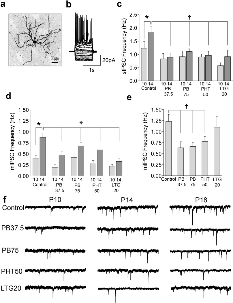
Methods: We used patch-clamp recordings to examine functional synaptic maturation in striatal medium spiny neurons from neonatal rats exposed to antiepileptic drugs with proapoptotic action (phenobarbital, phenytoin, lamotrigine) and without proapoptotic action (levetiracetam). Phenobarbital-exposed rats were also assessed for reversal learning at weaning.

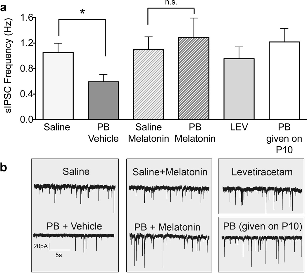
Results: Recordings from control animals revealed increased inhibitory and excitatory synaptic connectivity between postnatal day (P)10 and P18. This maturation was absent in rats exposed at P7 to a single dose of phenobarbital, phenytoin, or lamotrigine. Additionally, phenobarbital exposure impaired striatal-mediated behavior on P25. Neuroprotective pretreatment with melatonin, which prevents drug-induced neurodevelopmental apoptosis, prevented the drug-induced disruption in maturation. Levetiracetam was found not to disrupt synaptic development.

Interpretation: Our results provide the first evidence that exposure to antiepileptic drugs during a sensitive postnatal period impairs physiological maturation of synapses in neurons that survive the initial drug insult. These findings suggest a mechanism by which early life exposure to antiepileptic drugs can impact cognitive and behavioral outcomes, underscoring the need to identify therapies that control seizures without compromising synaptic maturation.


Figures

Figure 1
Figure 1
AED exposure disrupts IPSC development during the second postnatal week. (A) Representative confocal image of a biocytin-filled striatal medium spiny neurons (MSNs) with local dendritic arborization and spiny dendrites. Calibration bar, 30µm (B) Representative current clamp recording from a MSN, showing the characteristic repetitive, non-adapting firing pattern in response to a series of 1s hyperpolarizing and depolarizing current injections (10 pA steps) from a holding potential of −60 mV. (C) Quantification (mean±S.E.M.) of spontaneous inhibitory postsynaptic current (sIPSC) frequency at P10 and P14 showing the effect of P7 exposure to saline (control, n=28, n=32), phenobarbital (PB, 37.5 mg/kg, n=11, n=9), phenobarbital (PB, 75 mg/kg, n=9, n=23), phenytoin (PHT, 50 mg/kg, n=12, n=23), and lamotrigine (LTG, 20 mg/kg, n=11, n=11). (D) Quantification (mean±S.E.M.) of miniature inhibitory postsynaptic current (mIPSC) frequency at P10 and P14 showing effects of P7 exposure to saline (n=21, n=30), phenobarbital (37.5 mg/kg, n=6, n=6), phenobarbital (75 mg/kg, n=6, n=23), phenytoin (50 mg/kg, n=12, n=21), and lamotrigine (20 mg/kg, n=9, n=8) (E) Quantification (mean±S.E.M.) of mIPSC frequency at P18 showing effects of P7 exposure to saline (n=22), phenobarbital (37.5 mg/kg, n=6), phenobarbital (75 mg/kg, n=9), phenytoin (50 mg/kg, n=8), and lamotrigine (20 mg/kg, n=10). (F) Representative mIPSC traces from each group at P10, P14 and P18, following P7 treatment with saline, phenobarbital (75 mg/kg), phenobarbital (37.5 mg/kg), phenytoin (50 mg/kg), and lamotrigine (20 mg/kg). Data were analyzed by ANOVA with Fisher’s Least Significant Difference test for multiple comparisons. * indicates significantly different than P10 value within treatment. † Indicates significantly different than control at same age. See Supplemental Statistics Discussion for a complete discussion of ANOVA results.
Figure 2
Figure 2
AED exposure disrupts excitatory postsynaptic current development and alters spine morphology. (A) Quantification of glutamatergic (miniature excitatory postsynaptic currents, mEPSCs) development in striatal MSNs from P10 to P18, as a function of drug treatment, showing effects of P7 exposure to saline (n=19, n=29, n=21), phenobarbital (PB, 37.5 mg/kg, n=6, n=6, n=7), phenobarbital (PB, 75 mg/kg, n=7, n=19, n=8), phenytoin (PHT, 50 mg/kg, n=9, n=19, n=8), and lamotrigine (LTG, 20 mg/kg, n=6, n=9, n=19). Data were analyzed by ANOVA with Fisher's Least Significant Difference post-hoc test. * indicates significantly different than P10 value within treatment (P<0.05), † indicates significantly different than control at same age (P<0.05). (B1) Quantification of spine and (B2) filopodia number on P18 MSNs from animals exposed to saline (control, n=8 neurons) or phenobarbital (75mg/kg, n=5 neurons) on P7; while the number of spines did not differ between treatments (t-test, P=0.9246), the number of filopodia where significantly greater in phenobarbital-exposed animals (n=6 neurons per treatment, t-test, * = P<0.0001). (B3) Quantification (performed using ImageJ) of spine length (left y-axis) and (B4) spine width (right y-axis) in P18 MSNs from animals exposed to control (n=6 neurons) or PB (75mg/kg, n=6 neurons) on P7. (C) Representative confocal images of dendritic branch used for quantification of spines and filopodia. Only mature (mushroom shaped) spines were counted (black arrow). Filopodia were classified as long thin processes (grey arrow) and were not included in spine width or length measurements.
Figure 3
Figure 3
Treatments that avoid cellular toxicity do not disrupt development. Spontaneous inhibitory postsynaptic current (sIPSC) frequency, measured at P14, in medium spiny neurons (MSNs) from animals exposed on P7 to saline (n=24), phenobaribtal/vehicle (n=10), saline/melatonin (n=13), phenobarbital/melatonin (n=14), levetiracetam (n=9) or exposed on P10 to phenobarbital (n=16). (* = p<0.05, t-test with Bonferroni’s correction for multiple comparisons).

References

    1. Hauser WA. The prevalence and incidence of convulsive disorders in children. Epilepsia. 1994;35(Suppl 2):S1–S6. - PubMed
    1. Parisi P, Francia A, Vanacore N, et al. Psychomotor development and general movements in offspring of women with epilepsy and anticonvulsant therapy. Early Hum. Dev. 2003;74(2):97–108. [cited 2011 Jun 10] - PubMed
    1. Reinisch JM, Sanders SA, Mortensen EL, Rubin DB. In utero exposure to phenobarbital and intelligence deficits in adult men. JAMA. 1995;274(19):1518–1525. [cited 2011 Jun 10] - PubMed
    1. Farwell JR, Lee YJ, Hirtz DG, et al. Phenobarbital for febrile seizures--effects on intelligence and on seizure recurrence. N. Engl. J. Med. 1990;322(6):364–369. [cited 2011 Jun 10] - PubMed
    1. Ikonomidou C, Scheer I, Wilhelm T, et al. Brain morphology alterations in the basal ganglia and the hypothalamus following prenatal exposure to antiepileptic drugs. Eur. J. Paediatr. Neurol. 2007;11(5):297–301. - PubMed

Publication types

MeSH terms