. 2012 Aug;40(15):7207-18.
doi: 10.1093/nar/gks384. Epub 2012 May 11.

The FleQ protein from Pseudomonas aeruginosa functions as both a repressor and an activator to control gene expression from the pel operon promoter in response to c-di-GMP

Affiliations

The FleQ protein from Pseudomonas aeruginosa functions as both a repressor and an activator to control gene expression from the pel operon promoter in response to c-di-GMP

Claudine Baraquet et al. Nucleic Acids Res. 2012 Aug.

Abstract

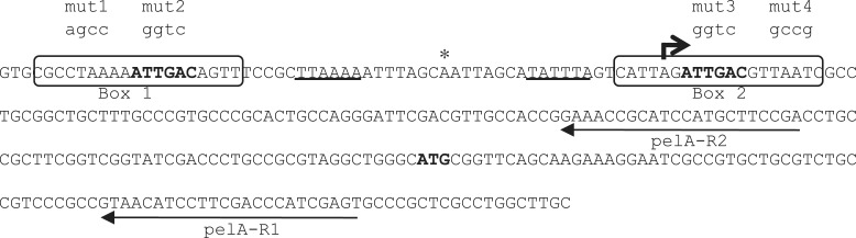
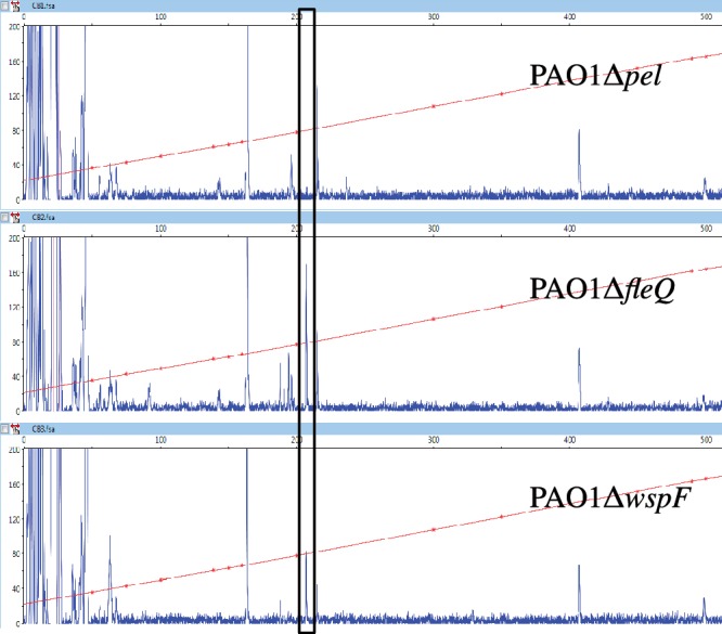
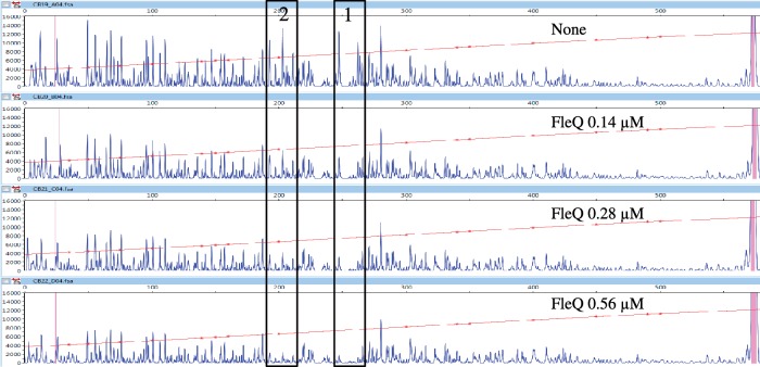
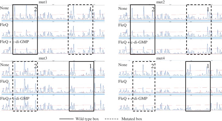
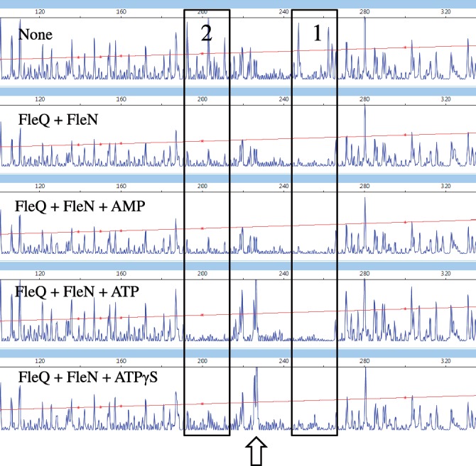
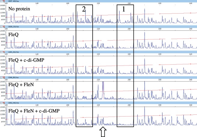
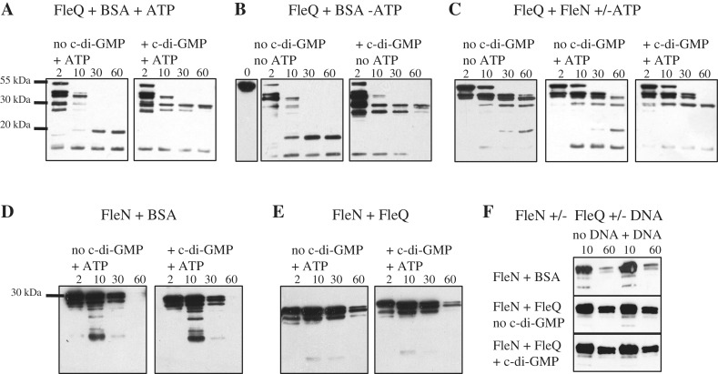
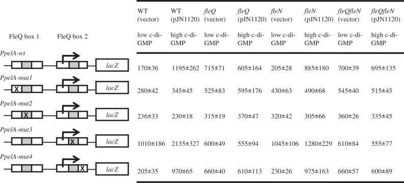
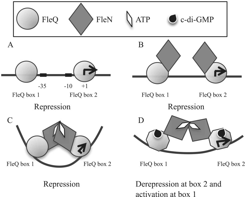
Bis-(3'-5')-cyclic dimeric guanosine monophosphate (c-di-GMP) modulates the transition between planktonic and biofilm life styles. In response to c-di-GMP, the enhancer binding protein FleQ from Pseudomonas aeruginosa derepresses the expression of Pel exopolysaccharide genes required for biofilm formation when a second protein, FleN is present. A model is that binding of c-di-GMP to FleQ induces its dissociation from the pelA promoter allowing RNA polymerase to access this site. To test this, we analyzed pelA DNA footprinting patterns with various combinations of FleQ, FleN and c-di-GMP, coupled to in vivo promoter activities. FleQ binds to two sites called box 1 and 2. FleN binds to FleQ bound at these sites causing the intervening DNA to bend. Binding of c-di-GMP to FleQ relieves the DNA distortion but FleQ remains bound to the two sites. Analysis of wild type and mutated versions of pelA-lacZ transcriptional fusions suggests that FleQ represses gene expression from box 2 and activates gene expression in response to c-di-GMP from box 1. The role of c-di-GMP is thus to convert FleQ from a repressor to an activator. The mechanism of action of FleQ is distinct from that of other bacterial transcription factors that both activate and repress gene expression from a single promoter.


Figures

Figure 1.
Figure 1.
Nucleotide sequence of the pel promoter region. The two FleQ binding sites are boxed and the repeated sequences are in bold. The identity of various promoter mutations is shown above the sequence. The transcription start site is indicated by a thick arrow and the putative −10 and −35 elements are underlined. The translation start site ATG is in bold. The asterisk indicates the location of the hypersensitive site. The two thin arrows indicate the two primers pelA-R1 and pelA-R2 used for primer extension and DNAse I footprinting experiments.
Figure 2.
Figure 2.
Identification of the transcription start site of pelA. Primer extension analysis of the pelA transcript was performed using a 6-FAM labeled primer (pelA-R1), which hybridizes to the RNA 49–73 bp downstream from the translation start site of pelA. Total RNA were isolated from PAO1Δpel, PAO1ΔfleQ and PAO1ΔwspF strains. The fluorescence intensity of FAM-labeled cDNA fragments (ordinate) is plotted against the sequence length of the fragment (abscissa). The horizontal line indicates the GS-500 internal size standard. The major transcript detected in strains PAO1ΔfleQ and PAO1ΔwspF but not detected in strain PAO1Δpel is boxed.
Figure 3.
Figure 3.
The pel promoter contains two FleQ boxes. 5′-FAM-labeled pel DNA (0.45 pmol) was incubated with or without FleQ (0.14, 0.28 or 0.56 μM, as indicated) and in the presence of 10 μM of ATP, then submitted to DNase I digestion (0.3 U) and analyzed by capillary electrophoresis. The fluorescence intensity of FAM-labeled DNA fragments (ordinate), in arbitrary units, is plotted against the sequence length of the fragment (abscissa), in bases, relative to the first base of the pelA-R1 primer. The whole electrophoregram is shown. The horizontal line indicates the GS-500 internal size standard. The two boxes labeled 1 and 2 indicate the two regions protected by FleQ.
Figure 4.
Figure 4.
Effects of mutations in the pelA promoter on the binding of FleQ. 5′-FAM-labeled pel DNA (0.45 pmol) carrying different nucleotide substitutions as indicated in Figure 1 was incubated in the presence of ATP (10 μM), with or without FleQ (0.28 μM) and in the presence or absence of c-di-GMP (100 μM), as indicated. The sequence length of the fragment, in bases, is relative to the first base of the pelA-R2 primer. Part of the electrophoregram is shown. The two boxes labeled 1 and 2 indicate the two regions of FleQ binding, the dashed line of the box indicates which box is mutated.
Figure 5.
Figure 5.
FleN causes the pel promoter to bend in conjunction with FleQ and ATP or ATPγS. Analysis of the 5′-FAM-labeled pel DNA (0.45 pmol) incubated with FleQ alone or with FleN at equimolar concentrations (0.28 μM) and in the presence and absence of ATP (10 μM), AMP (10 μM) or ATPγS (10 μM). The sequence length of the fragment (abscissa), in bases, is relative to the first base of the pelA-R1 primer. The two FleQ binding sites are boxed and the hypersensitive site is indicated by an arrow.
Figure 6.
Figure 6.
Effect of c-di-GMP on FleQ and FleN binding to the pel promoter. Analysis of the 5′-FAM-labeled pel DNA (0.45 pmol) incubated with FleQ alone or with FleN at equimolar concentrations (0.28 μM), in the presence of ATP (10 μM) and in the presence or absence of c-di-GMP (100 μM). The sequence length of the fragment (abscissa), in bases, is relative to the first base of the pelA-R1 primer. The two FleQ binding sites are boxed and the hypersensitive site is indicated by an arrow.
Figure 7.
Figure 7.
Effects of c-di-GMP and ATP on the kinetics of trypsin digestion of FleQ alone or FleQ and FleN. FleQ and BSA (A and B), FleN and BSA (D) or FleQ and FleN (C, E and F) were incubated in the presence or absence of c-di-GMP (1 mM), ATP (100 μM) or a DNA fragment encompassing the two FleQ binding sites (3.2 μM), as indicated and then subjected to proteolysis with trypsin. Times of incubation are indicated above the gel. Digestion products were separated by SDS–PAGE, transferred to a nitrocellulose membrane and detected with anti-FleQ antibodies (A, B and C) or with anti-FleN antibodies (D, E and F).
Figure 8.
Figure 8.
FleQ acts as a repressor in the absence of c-di-GMP and as an activator in the presence of c-di-GMP. β-Galactosidase activities of wild type or mutated PpelA-lacZ fusions, as indicated on the left of the diagram, in PAO1ΔpelΔpsl (WT), PAO1ΔpelΔpslΔfleQ (fleQ), PAO1ΔpelΔpslΔfleN (fleN) or PAO1ΔpelΔpslΔfleQΔfleN (fleQfleN) strains carrying pJN105 (vector control) or pJN1120 (allowing the overexpression of the DGC encoded by PA1120). The white boxes indicate the FleQ boxes, the grey boxes, the repeated sequences observed in the FleQ boxes. Mutations in either boxes are indicated by an X and the transcription start site is indicated by an arrow. The β-galactosidase values are indicated on the right and expressed as means ± standard deviation.
Figure 9.
Figure 9.
Model of pel regulation. FleQ binds to two FleQ boxes on the pel promoter (A). FleQ interacts with FleN in the absence of ATP (B) as well as in the presence of ATP, but in this case it induces a distortion of pel DNA (C). We propose that FleN forms a bridge between two FleQ bound to their binding sites. The binding of FleQ to FleQ box 2 is essential for repression. The binding of c-di-GMP to FleQ induces a conformational change of FleQ, probably propagated through FleN, which induces the relief of pel distortion and leads to pel expression (D). The binding of FleQ to FleQ box 1 is essential for activation.

References

    1. Hickman JW, Tifrea DF, Harwood CS. A chemosensory system that regulates biofilm formation through modulation of cyclic diguanylate levels. Proc. Natl. Acad. Sci. USA. 2005;102:14422–14427. - PMC - PubMed
    1. Simm R, Morr M, Kader A, Nimtz M, Romling U. GGDEF and EAL domains inversely regulate cyclic di-GMP levels and transition from sessility to motility. Mol. Microbiol. 2004;53:1123–1134. - PubMed
    1. Tischler AD, Camilli A. Cyclic diguanylate (c-di-GMP) regulates Vibrio cholerae biofilm formation. Mol. Microbiol. 2004;53:857–869. - PMC - PubMed
    1. Wolfe AJ, Visick KL. Get the message out: cyclic-Di-GMP regulates multiple levels of flagellum-based motility. J. Bacteriol. 2008;190:463–475. - PMC - PubMed
    1. Thormann KM, Duttler S, Saville RM, Hyodo M, Shukla S, Hayakawa Y, Spormann AM. Control of formation and cellular detachment from Shewanella oneidensis MR-1 biofilms by cyclic di-GMP. J. Bacteriol. 2006;188:2681–2691. - PMC - PubMed

Publication types

MeSH terms