

Inverse cutting of posterior lamellar corneal grafts by a femtosecond laser
- PMID: 22582107
- PMCID: PMC3349952
- DOI: 10.2174/1874364101206010019
Inverse cutting of posterior lamellar corneal grafts by a femtosecond laser
Abstract
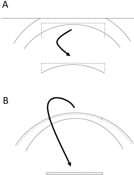
Purpose: Posterior lamellar grafting of the cornea has become the preferred technique for treatment of corneal endothelial dysfunction. Posterior lamellar grafts are usually cut by a micro-keratome or a femto-second laser after the epithelial side of the donor cornea has been applanated. This approach often results in variable central graft thickness in different grafts and an increase in graft thickness towards the periphery in every graft. The purpose of this study was to evaluate if posterior lamellar grafts can be prepared from the endothelial side by a femto-second laser, resulting in reproducible, thin grafts of even thickness.
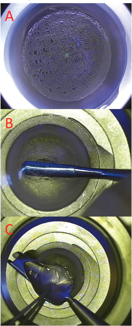
Methods: A CZM 500 kHz Visumax femto-second laser was used. Organ cultured donor grafts were mounted in an artifical anterior chamber with the endothelial side up and out. Posterior grafts of 7.8 mm diameter and 130 micron thickness were prepared by femto-second laser cutting. A standard DSAEK procedure was performed in 10 patients with Fuchs endothelial dystrophy. Patients were followed-up regularly and evaluated by measurement of complications, visual acuity, corneal thickness (Pentacam HR), and endothelial cell density.
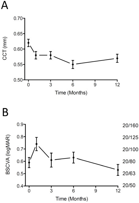
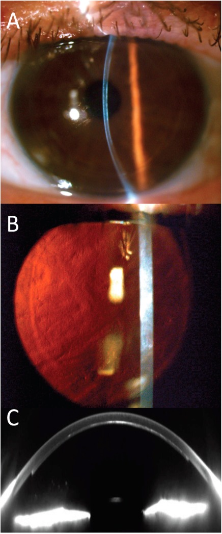
Results: Femto-laser cutting of grafts and surgery was uncomplicated. Rebubbling was necessary in 5 of 10 cases (normally only in 1 of 20 cases). All grafts were attached and cleared up during the first few weeks. After six months, the average visual acuity was 0.30 (range: 0.16 to 0.50), corneal thickness was 0.58 mm (range 0.51 to 0.63), and endothelial cell density was 1.570 per sq. mm (range: 1.400 to 2.000 cells per sq. mm). The grafts were of uniform thickness, but substantial interface haze was present in most grafts.
Conclusions: Posterior lamellar corneal grafts can be prepared from the endothelial side using a femto-second laser. All grafts were clear after 6 months with satisfying endothelial cell counts. Poor visual acuity caused by interface scatter was observed in most patients. Femto-second laser cutting parameters needs to be optimised to enable smooth cutting in the posterior stroma.
Keywords: Corneal grafts; DSAEK.; Femto-laser; cornea.
Figures
References
-
- Chen ES, Terry MA, Shamie N, et al. Descemet-stripping automated endothelial keratoplasty: six-month results in a prospective study of 100 eyes. Cornea. 2008;27:514–20. - PubMed
-
- Jun B, Kuo AN, Afshari NA, et al. Refractive change after Descemet stripping automated endothelial keratoplasty surgery and its correlation with graft thickness and diameter. Cornea. 2009;28:19–23. - PubMed
-
- Cheng YY, Schouten JS, Tahzib NG, et al. Efficacy and safety of femtosecond laser-assisted corneal endothelial keratoplasty a randomized multicenter clinical trial. Transplantation. 2009;88(11):1294–302. - PubMed
-
- Hjortdal J, Ehlers N, et al. Descemet's stripping automated endothelial keratoplasty and penetrating keratoplasty for Fuchs' endothelial dystrophy. Acta Ophthalmol. 2009;87(3):310–4. - PubMed
LinkOut - more resources
Full Text Sources
Other Literature Sources