. 2012 Jul 1;303(1):L1-L11.
doi: 10.1152/ajplung.00050.2012. Epub 2012 May 11.

Osteopontin is an endogenous modulator of the constitutively activated phenotype of pulmonary adventitial fibroblasts in hypoxic pulmonary hypertension

Affiliations

Osteopontin is an endogenous modulator of the constitutively activated phenotype of pulmonary adventitial fibroblasts in hypoxic pulmonary hypertension

Adil Anwar et al. Am J Physiol Lung Cell Mol Physiol. .

Abstract

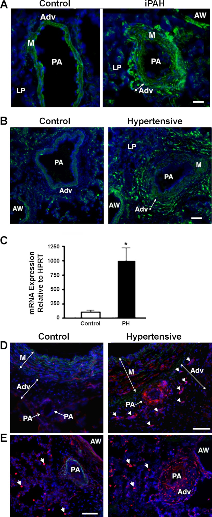
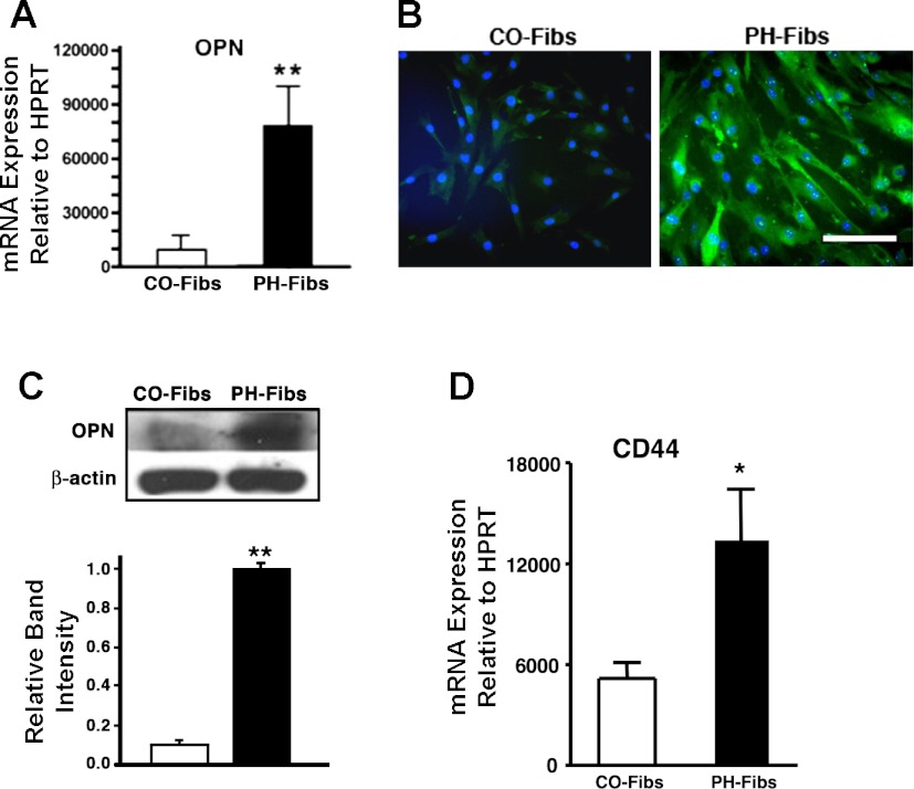
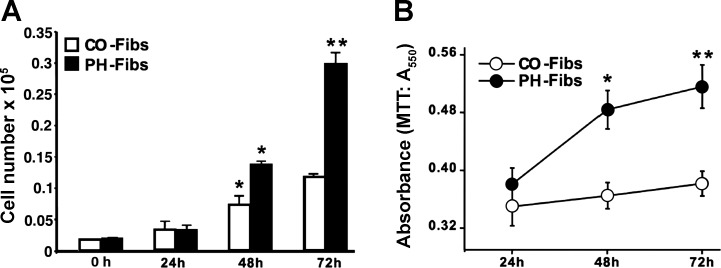
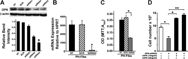
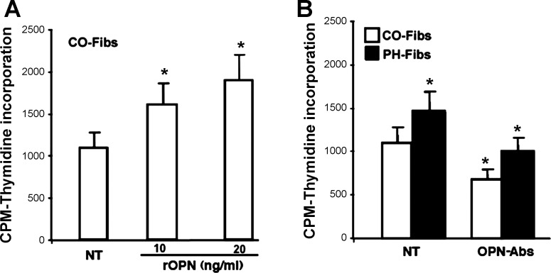
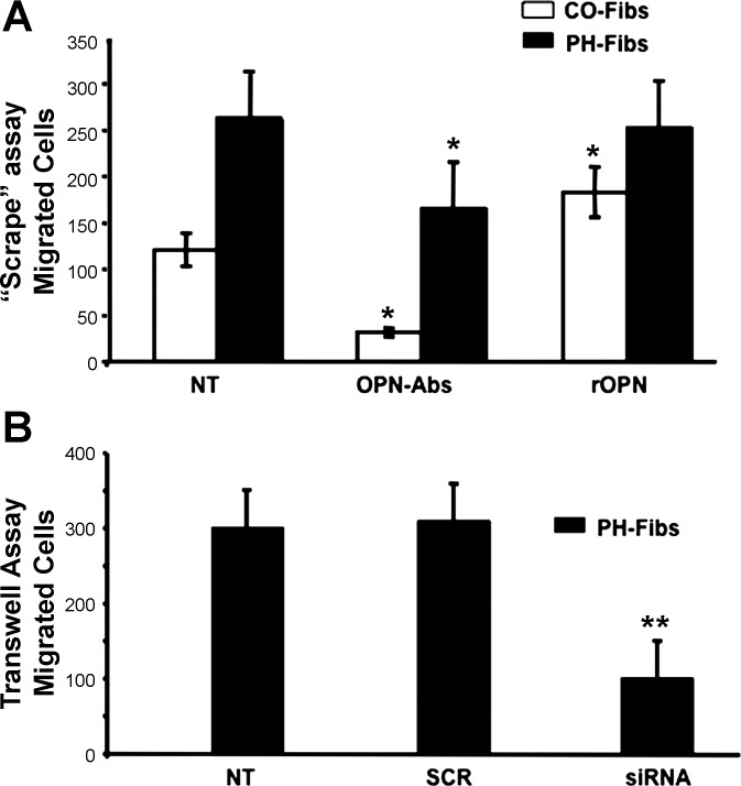
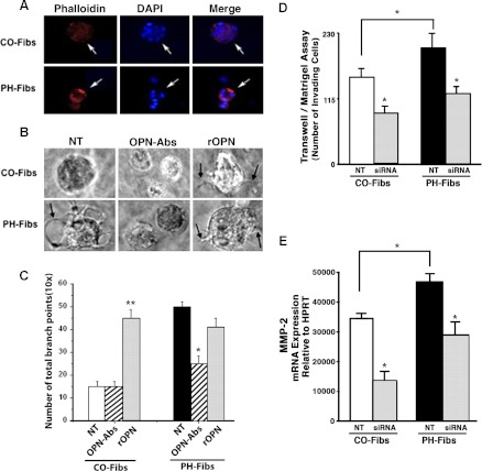
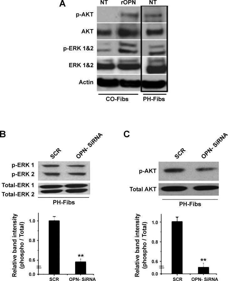
Increased cell proliferation and migration, of several cell types are key components of vascular remodeling observed in pulmonary hypertension (PH). Our previous data demonstrate that adventitial fibroblasts isolated from pulmonary arteries of chronically hypoxic hypertensive calves (termed PH-Fibs) exhibit a "constitutively activated" phenotype characterized by high proliferative and migratory potential. Osteopontin (OPN) has been shown to promote several cellular activities including growth and migration in cancer cells. We thus tested the hypothesis that elevated OPN expression confers the "activated" highly proproliferative and promigratory/invasive phenotype of PH-Fibs. Our results demonstrate that, both in vivo and ex vivo, PH-Fibs exhibited increased expression of OPN, as well as its cognate receptors, α(V)β(3) and CD44, compared with control fibroblasts (CO-Fibs). Augmented OPN expression in PH-Fibs corresponded to their high proliferative, migratory, and invasive properties and constitutive activation of ERK1/2 and AKT signaling. OPN silencing via small interfering RNA or sequestering OPN production by specific antibodies led to decreased proliferation, migration, invasion, and attenuated ERK1/2, AKT phosphorylation in PH-Fibs. Furthermore, increasing OPN levels in CO-Fibs via recombinant OPN resulted in significant increases in their proliferative, migratory, and invasive capabilities to the levels resembling those of PH-Fibs. Thus our data suggest OPN as an essential contributor to the activated (highly proliferative, migratory, and proinvasive) phenotype of pulmonary adventitial fibroblasts in hypoxic PH.


Figures

Fig. 1.
Fig. 1.
Osteopontin (OPN) expression is upregulated in hypoxia-induced pulmonary hypertension (PH) (in vivo assessment). AC: pulmonary hypertension-associated vascular remodeling is characterized by upregulation of OPN. A: in human subjects with idiopathic pulmonary arterial hypertension (iPAH, right), marked upregulation of OPN expression is observed in vascular adventitia, compared with control specimens (Control, left). B: upregulation of OPN protein expression is observed in vascular adventitia (Adv) of chronically hypoxic hypertensive calves (right) compared with normotensive controls (left). Lung cryosections in A and B were labeled with OPN-specific antibodies (green fluorescence) and counterstained with 4,6-diamidino-2-phenylindole (DAPI) (cell nuclei, blue). C: OPN mRNA expression is upregulated in whole lung extracts from neonatal calves with chronic hypoxia-induced PH compared with age-matched control calves. HPRT, hypoxanthine-xanthine phosphoribosyl transferase. D and E: upregulated expression of the OPN receptors αVβ3 integrin (D) and CD44 (E) is observed in pulmonary vascular adventitia of chronically hypertensive calves (right) compared with normoxic controls (left). In chronically hypoxic hypertensive calves, adventitia of a smaller-size vessel (PA with an arrow) as well as cells in the adventitia (arrowheads) of a larger elastic intralobar pulmonary artery (PA) demonstrate intense positive staining for αVβ3 integrin (red fluorescence). Vascular media (M) also show increase in reactivity. In control normoxic animals, both vascular adventitia and media display minimal expression of OPN. Number of specimens analyzed: CO and PH calves (n = 7, each group); human subjects (controls, n = 4; iPAH, n = 5). Scale bars in A and C = 50 μM; in D and E = 100 μM. AW, airway; LP, lung parenchyma.
Fig. 2.
Fig. 2.
Pulmonary adventitial fibroblasts isolated from severely hypertensive calves (PH-Fibs) express markedly higher levels of OPN and OPN receptor CD44 than control fibroblasts (CO-Fibs). A: quantitative real-time PCR demonstrates that PH-Fibs express 8-fold higher OPN mRNA levels than CO-Fibs. Data presented are relative OPN transcript abundance to HPRT. B and C: at the protein level, as shown by immunofluorescent staining (B) and Western blotting (C), OPN expression levels are markedly higher in PH-Fibs compared with CO-Fibs. Scale bar in B = 50 μM. C: Western blotting (top) was quantified by densitometric scanning analysis (bottom); data are representative of at least 3 independent experiments. D: RT-PCR analysis demonstrates that PH-Fibs express OPN receptor CD44 at levels 2.58 ± 0.59-fold higher than those in CO-Fibs. Data presented are relative OPN transcript abundance to HPRT. *P < 0.05, **P < 0.01.
Fig. 3.
Fig. 3.
PH-Fibs proliferate at markedly higher rates than CO-Fibs. A and B: cell proliferation under serum-free conditions was assessed by cell number counts (A) and 3-(4,5-dimethylthiozol-2-yl)-2,5-diphenyltetrazolium bromide (MTT) assay (B) over a 72-h period (see methods). Results are expressed as means ± SE; *P < 0.05; **P < 0.01.
Fig. 4.
Fig. 4.
Silencing of OPN by small interfering RNA (siRNA) in PH-Fibs results in attenuated cell proliferation. A and B: different siRNAs (siRNA 1–3) were tested for their ability to block OPN protein expression in PH-Fibs and siRNA-3 was chosen as the best one. As shown by both Western blotting (A) and real-time PCR analysis (B), treatment of PH-Fibs with siRNA-3 (50 nM, 48 h for RT-PCR, 72 h for Western blotting) mediated significant downregulation of OPN expression. C: siRNA3-mediated OPN silencing in PH-Fibs resulted in decreased cell proliferation as shown by MTT assay (72-h time point). D: addition of recombinant OPN (rOPN; 10 or 20 ng/ml) to siRNA-treated PH-Fibs restored and even elevated their proliferative potential, as determined by cell count assessment. Data shown are representative of at least 3 independent experiments. NT, not treated; SCR, treated with scrambled RNA; siRNA 1–3, treated with siRNA 1, 2, or 3. *P < 0.05; **P < 0.01.
Fig. 5.
Fig. 5.
Modulation of OPN expression in PH- and CO-Fibs results in alterations of their proproliferative capabilities. A: overexpression of OPN in CO-Fibs by treatment with rOPN (10 or 20 ng/ml, 24 h) resulted in augmented DNA synthesis. B: treatment of PH-Fibs and CO-Fibs with OPN-specific antibodies (7.5 μg/ml, 24 h) resulted in significantly attenuated DNA synthesis in both cell types. OPN-Abs, treated with OPN-specific antibodies. Number of cell populations analyzed: PH-Fibs, n = 5; CO-Fibs, n = 4. Results are expressed as mean ± SE; *P < 0.05.
Fig. 6.
Fig. 6.
Elevated migratory capabilities of PH-Fibs are attenuated upon downregulation of OPN expression. A: in a “scrape” assay (performed with addition of hydroxyurea to inhibit cell proliferation; see methods), sequestering OPN production by treatment with OPN-specific antibodies (7.5 μg/ml, 48 h) resulted in significantly attenuated migratory capabilities of both CO- and PH-Fibs (OPN-Abs). Addition of rOPN augmented migratory potential of CO-Fibs; however, it did not affect PH-Fibs. Number of cell populations analyzed: PH-Fibs, n = 5; CO-Fibs, n = 4. B: in a Boyden Transwell chamber assay under serum-free conditions, OPN knockdown by siRNA-3 (50 nM, 48 h) resulted in markedly attenuated migration of PH-Fibs. Data represent mean ± SE from 3 independent experiments. Number of PH-Fibs populations analyzed, n = 3; *P < 0.01 vs. NT cells.
Fig. 7.
Fig. 7.
OPN expression levels modulate proinvasive capabilities of PH- and CO-Fibs in 3D Matrigel assays. A: cells were plated onto growth factor-reduced Matrigel in serum-free medium. After 9 days, immunostaining for actin (via Alexa-594-conjugated phalloidin, red) to define cell morphology, and for cell nuclei (DAPI, blue) was performed. B: in 3D growth factor-reduced Matrigel cultures PH-Fibs generated visible filopodia-like structures, a hallmark of cell invasion into Matrigel (arrow in NT image), as opposed to CO-Fibs. In the presence of OPN-specific antibodies (7.5 μg/ml), generation of filopodia in PH-Fibs was inhibited. Addition of rOPN (20 ng/ml) resulted in generation of filopodia (arrows) not only in PH-Fibs, but even in CO-Fibs that did not occur under NT conditions. C: quantification of filopodia-like structures (the number of branch points) was performed via ibidi software analysis: http://www.ibidi.com (Munich, Germany). **P < 0.01, *P < 0.05. D: in Matrigel-coated Boyden chamber assay of cell invasion (see methods), siRNA3-mediated knockdown of OPN expression resulted in reduced invasion through Matrigel (used in a growth factor-reduced form) in both CO- and PH-Fibs. Data represent mean ± SE from 3 different experiments. *P < 0.05 vs. NT cells. E: under NT conditions, MMP-2 expression in PH-Fibs was significantly higher than in CO-Fibs. Treatment of both CO- and PH-Fibs with OPN-siRNA (50 nM, 48 h) resulted in significant decrease of MMP-2 expression (6-fold in CO-Fibs and 4-fold in PH-Fibs) compared with respective NT cells.
Fig. 8.
Fig. 8.
OPN expression controls activation state of ERK1/2 and AKT signaling pathways in CO- and PH-Fibs. A: under serum-free conditions, PH-Fibs exhibited constitutively activated [phosphorylated (p-)] ERK1/2 and AKT signaling pathways (PH-Fibs, NT lane). In contrast, under serum-free conditions, CO-Fibs exhibited low- to not- phosphorylated states of ERK1/2 and AKT, respectively (CO-Fibs, NT lane). Treatment of CO-Fibs with rOPN (20 ng/ml, 6 h) resulted in profound activation (phosphorylation) of both ERK1/2 and AKT signaling pathways (CO-Fibs, rOPN lane). B and C: treatment of PH-Fibs, maintained under serum-free conditions, with OPN-siRNA-3 (50 nM, 72 h) resulted in significant downregulation of both phospho-ERK1/2 (B) and phospho-AKT (C) compared with scrambled oligos (SCR). Data represent mean ± SE from 3 different experiments. **P < 0.01.

References

    1. Angelucci A, Festuccia C, Gravina GL, Muzi P, Bonghi L, Vicentini C, Bologna M. Osteopontin enhances the cell proliferation induced by the epidermal growth factor in human prostate cancer cells. Prostate 59: 157–166, 2004 - PubMed
    1. Anwar A, Dehn D, Siegel D, Kepa JK, Tang LJ, Pietenpol JA, Ross D. Interaction of human NAD(P)H:quinone oxidoreductase 1 (NQO1) with the tumor suppressor protein p53 in cells and cell-free systems. J Biol Chem 278: 10368–10373, 2003 - PubMed
    1. Archer S, Rich S. Primary pulmonary hypertension: a vascular biology and translational research “Work in progress”. Circulation 102: 2781–2791, 2000 - PubMed
    1. Bhowmick NA, Neilson EG, Moses HL. Stromal fibroblasts in cancer initiation and progression. Nature 432: 332–337, 2004 - PMC - PubMed
    1. Burke DL, Frid MG, Kunrath CL, Karoor V, Anwar A, Wagner BD, Strassheim D, Stenmark KR. Sustained hypoxia promotes the development of a pulmonary artery-specific chronic inflammatory microenvironment. Am J Physiol Lung Cell Mol Physiol 297: L238–L250, 2009 - PMC - PubMed

Publication types

MeSH terms